966C | pdb_0000966c

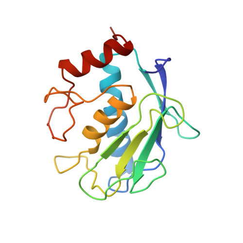
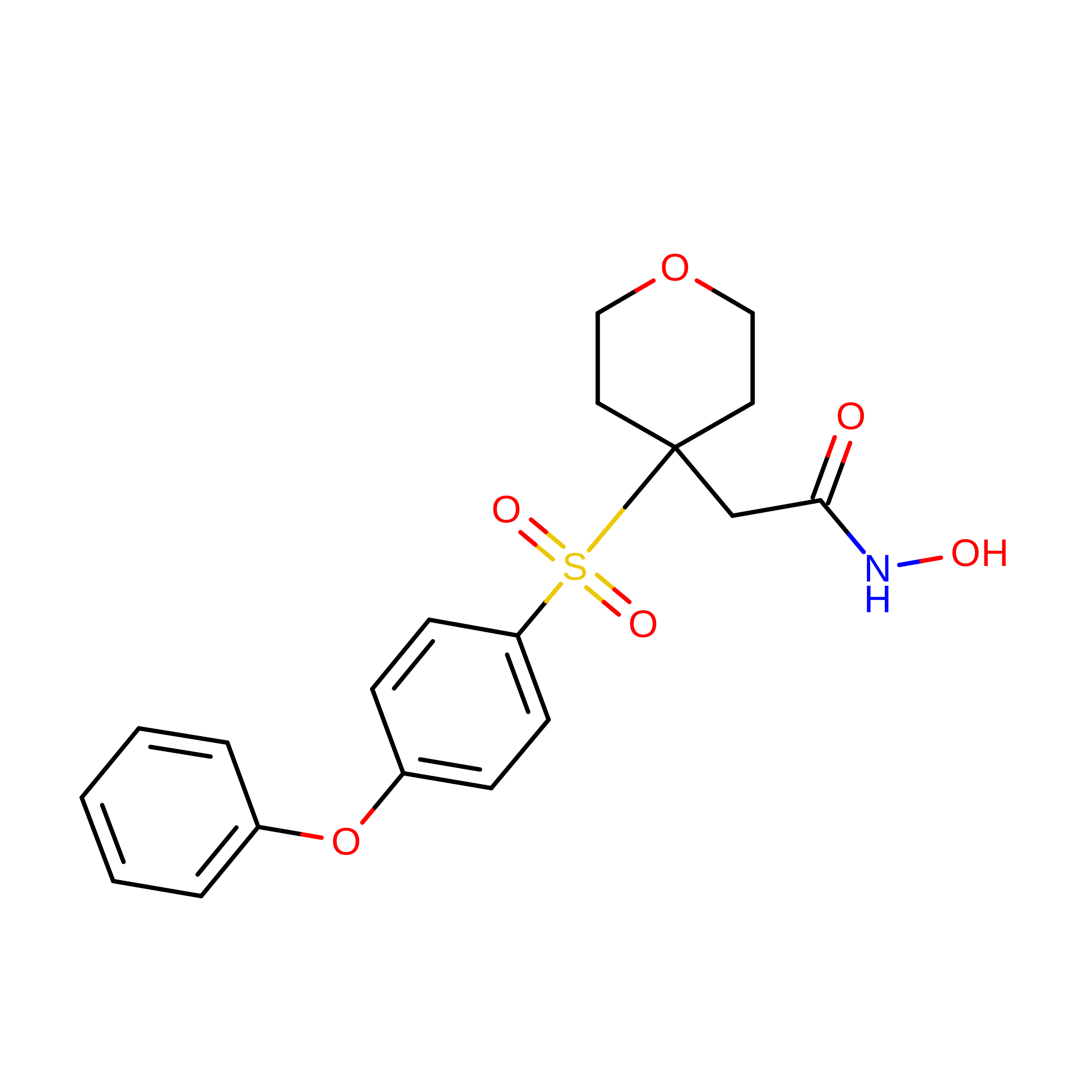
CRYSTAL STRUCTURE OF FIBROBLAST COLLAGENASE-1 COMPLEXED TO A DIPHENYL-ETHER SULPHONE BASED HYDROXAMIC ACID


Experimental Data Snapshot

  • Method: X-RAY DIFFRACTION
  • Resolution: 1.90 Å
  • R-Value Free: 
    0.330 (Depositor) 
  • R-Value Work: 
    0.218 (Depositor) 
  • R-Value Observed: 
    0.218 (Depositor) 

wwPDB Validation 3D Report Full Report

Validation slider image for 966C

This is version 1.4 of the entry. See complete history. 

Literature

Crystal structures of MMP-1 and -13 reveal the structural basis for selectivity of collagenase inhibitors.

Lovejoy, B., Welch, A.R., Carr, S., Luong, C., Broka, C., Hendricks, R.T., Campbell, J.A., Walker, K.A., Martin, R., Van Wart, H., Browner, M.F.

(1999) Nat Struct Biol 6: 217-221

  • DOI: https://doi.org/10.1038/6657
  • Primary Citation Related Structures: 
    456C, 830C, 966C

  • PubMed Abstract: 

    The X-ray crystal structures of the catalytic domain of human collagenase-3 (MMP-13) and collagenase-1 (MMP-1) with bound inhibitors provides a basis for understanding the selectivity profile of a novel series of matrix metalloprotease (MMP) inhibitors. Differences in the relative size and shape of the MMP S1' pockets suggest that this pocket is a critical determinant of MMP inhibitor selectivity. The collagenase-3 S1' pocket is long and open, easily accommodating large P1' groups, such as diphenylether. In contrast, the collagenase-1 S1' pocket must undergo a conformational change to accommodate comparable P1' groups. The selectivity of the diphenylether series of inhibitors for collagenase-3 is largely determined by their affinity for the preformed S1' pocket of collagenase-3, as compared to the induced fit in collagenase-1.


  • Organizational Affiliation: 
    • Inflammatory Diseases Unit, Roche Bioscience, Palo Alto, California 94304, USA.

Macromolecule Content 

  • Total Structure Weight: 18.26 kDa 
  • Atom Count: 1,405 
  • Modeled Residue Count: 157 
  • Deposited Residue Count: 157 
  • Unique protein chains: 1

Macromolecules

Find similar proteins by:|  3D Structure
Entity ID: 1
MoleculeChains  Sequence LengthOrganismDetailsImage
MMP-1157Homo sapiensMutation(s): 0 
EC: 3.4.24 (PDB Primary Data), 3.4.24.7 (UniProt)
UniProt & NIH Common Fund Data Resources
Find proteins for P03956 (Homo sapiens)
Explore P03956 
Go to UniProtKB:  P03956
PHAROS:  P03956
GTEx:  ENSG00000196611 
Entity Groups
Sequence Clusters30% Identity50% Identity70% Identity90% Identity95% Identity100% Identity
UniProt GroupP03956
Sequence Annotations
Expand
Reference Sequence

Experimental Data & Validation

Experimental Data

  • Method: X-RAY DIFFRACTION
  • Resolution: 1.90 Å
  • R-Value Free:  0.330 (Depositor) 
  • R-Value Work:  0.218 (Depositor) 
  • R-Value Observed: 0.218 (Depositor) 
Space Group: P 21 21 21
Unit Cell:
Length ( Å )Angle ( ˚ )
a = 38.48α = 90
b = 56.52β = 90
c = 74.23γ = 90
Software Package:
Software NamePurpose
X-PLORmodel building
X-PLORrefinement
DENZOdata reduction
SCALEPACKdata scaling
X-PLORphasing

Structure Validation

View Full Validation Report


Entry History 

Deposition Data

Revision History  (Full details and data files)

  • Version 1.0: 1999-08-07
    Type: Initial release
  • Version 1.1: 2008-03-03
    Changes: Version format compliance
  • Version 1.2: 2011-07-13
    Changes: Version format compliance
  • Version 1.3: 2018-03-07
    Changes: Data collection, Other
  • Version 1.4: 2024-02-14
    Changes: Data collection, Database references, Derived calculations