830C

COLLAGENASE-3 (MMP-13) COMPLEXED TO A SULPHONE-BASED HYDROXAMIC ACID


Experimental Data Snapshot

  • Method: X-RAY DIFFRACTION
  • Resolution: 1.60 Å
  • R-Value Free: 0.266 
  • R-Value Work: 0.208 
  • R-Value Observed: 0.208 

wwPDB Validation   3D Report Full Report


This is version 1.3 of the entry. See complete history


Literature

Crystal structures of MMP-1 and -13 reveal the structural basis for selectivity of collagenase inhibitors.

Lovejoy, B.Welch, A.R.Carr, S.Luong, C.Broka, C.Hendricks, R.T.Campbell, J.A.Walker, K.A.Martin, R.Van Wart, H.Browner, M.F.

(1999) Nat Struct Biol 6: 217-221

  • DOI: https://doi.org/10.1038/6657
  • Primary Citation of Related Structures:  
    456C, 830C, 966C

  • PubMed Abstract: 

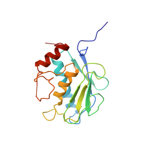
    The X-ray crystal structures of the catalytic domain of human collagenase-3 (MMP-13) and collagenase-1 (MMP-1) with bound inhibitors provides a basis for understanding the selectivity profile of a novel series of matrix metalloprotease (MMP) inhibitors. Differences in the relative size and shape of the MMP S1' pockets suggest that this pocket is a critical determinant of MMP inhibitor selectivity. The collagenase-3 S1' pocket is long and open, easily accommodating large P1' groups, such as diphenylether. In contrast, the collagenase-1 S1' pocket must undergo a conformational change to accommodate comparable P1' groups. The selectivity of the diphenylether series of inhibitors for collagenase-3 is largely determined by their affinity for the preformed S1' pocket of collagenase-3, as compared to the induced fit in collagenase-1.


  • Organizational Affiliation

    Inflammatory Diseases Unit, Roche Bioscience, Palo Alto, California 94304, USA.


Macromolecules
Find similar proteins by:  (by identity cutoff)  |  3D Structure
Entity ID: 1
MoleculeChains Sequence LengthOrganismDetailsImage
MMP-13
A, B
168Homo sapiensMutation(s): 0 
EC: 3.4.24
UniProt & NIH Common Fund Data Resources
Find proteins for P45452 (Homo sapiens)
Explore P45452 
Go to UniProtKB:  P45452
PHAROS:  P45452
GTEx:  ENSG00000137745 
Entity Groups  
Sequence Clusters30% Identity50% Identity70% Identity90% Identity95% Identity100% Identity
UniProt GroupP45452
Sequence Annotations
Expand
  • Reference Sequence
Small Molecules
Binding Affinity Annotations 
IDSourceBinding Affinity
RS1 BindingDB:  830C Ki: min: 0.28, max: 0.6 (nM) from 3 assay(s)
IC50: min: 1.00e-2, max: 34 (nM) from 5 assay(s)
PDBBind:  830C Ki: 0.52 (nM) from 1 assay(s)
Binding MOAD:  830C Ki: 0.52 (nM) from 1 assay(s)
Experimental Data & Validation

Experimental Data

  • Method: X-RAY DIFFRACTION
  • Resolution: 1.60 Å
  • R-Value Free: 0.266 
  • R-Value Work: 0.208 
  • R-Value Observed: 0.208 
  • Space Group: C 1 2 1
Unit Cell:
Length ( Å )Angle ( ˚ )
a = 136.072α = 90
b = 36.098β = 131.06
c = 96.368γ = 90
Software Package:
Software NamePurpose
X-PLORmodel building
X-PLORrefinement
MOSFLMdata reduction
CCP4data scaling
X-PLORphasing

Structure Validation

View Full Validation Report


Entry History 

Deposition Data

Revision History  (Full details and data files)

  • Version 1.0: 1999-08-06
    Type: Initial release
  • Version 1.1: 2008-03-03
    Changes: Version format compliance
  • Version 1.2: 2011-07-13
    Changes: Version format compliance
  • Version 1.3: 2023-08-09
    Changes: Database references, Derived calculations, Other, Refinement description