8XMN | pdb_00008xmn

Voltage-gated sodium channel Nav1.7 variant M2


Experimental Data Snapshot

  • Method: ELECTRON MICROSCOPY
  • Resolution: 3.37 Å
  • Aggregation State: PARTICLE 
  • Reconstruction Method: SINGLE PARTICLE 

wwPDB Validation 3D Report Full Report

Validation slider image for 8XMN

This is version 1.1 of the entry. See complete history

Literature

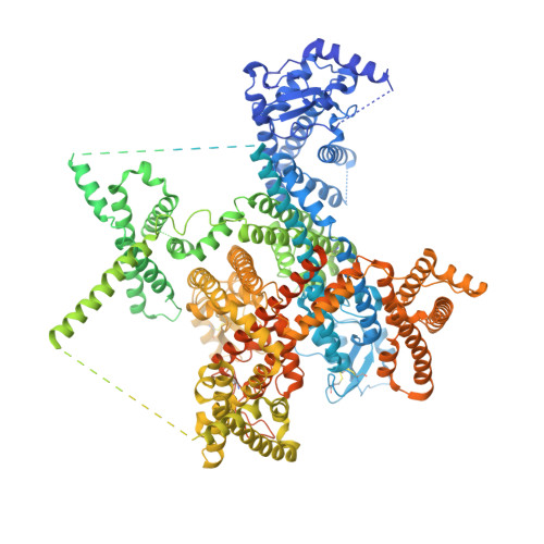
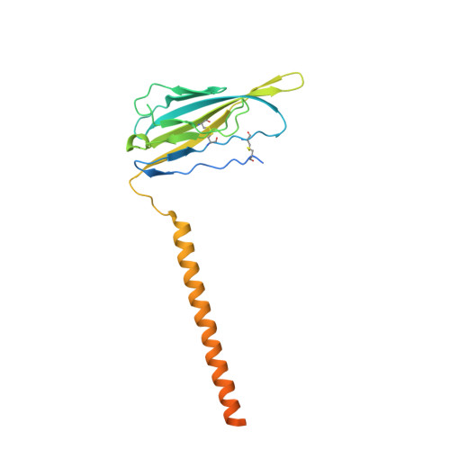
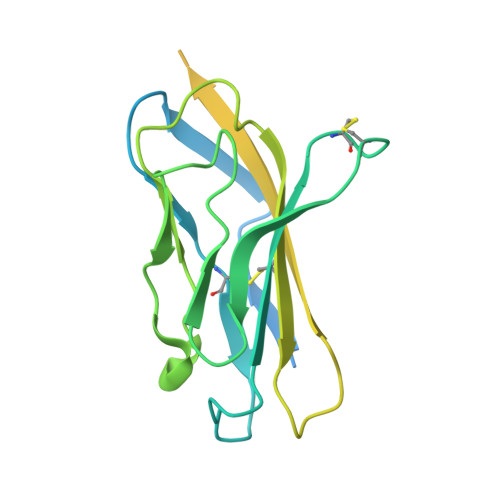
Dissection of the structure-function relationship of Na v channels.

Li, Z.Wu, Q.Huang, G.Jin, X.Li, J.Pan, X.Yan, N.

(2024) Proc Natl Acad Sci U S A 121: e2322899121-e2322899121

  • DOI: https://doi.org/10.1073/pnas.2322899121
  • Primary Citation Related Structures: 
    8XMM, 8XMN, 8XMO

  • PubMed Abstract: 

    Voltage-gated sodium channels (Na v ) undergo conformational shifts in response to membrane potential changes, a mechanism known as the electromechanical coupling. To delineate the structure-function relationship of human Na v channels, we have performed systematic structural analysis using human Na v 1.7 as a prototype. Guided by the structural differences between wild-type (WT) Na v 1.7 and an eleven mutation-containing variant, designated Na v 1.7-M11, we generated three additional intermediate mutants and solved their structures at overall resolutions of 2.9-3.4 Å. The mutant with nine-point mutations in the pore domain (PD), named Na v 1.7-M9, has a reduced cavity volume and a sealed gate, with all voltage-sensing domains (VSDs) remaining up. Structural comparison of WT and Na v 1.7-M9 pinpoints two residues that may be critical to the tightening of the PD. However, the variant containing these two mutations, Na v 1.7-M2, or even in combination with two additional mutations in the VSDs, named Na v 1.7-M4, failed to tighten the PD. Our structural analysis reveals a tendency of PD contraction correlated with the right shift of the static inactivation I-V curves. We predict that the channel in the resting state should have a "tight" PD with down VSDs.


  • Organizational Affiliation
    • Beijing Frontier Research Center for Biological Structures, State Key Laboratory of Membrane Biology, Tsinghua-Peking Joint Center for Life Sciences, School of Life Sciences, Tsinghua University, Beijing 100084, China.

Macromolecule Content 

  • Total Structure Weight: 290.72 kDa 
  • Atom Count: 13,110 
  • Modeled Residue Count: 1,569 
  • Deposited Residue Count: 2,488 
  • Unique protein chains: 3

Macromolecules

Find similar proteins by:|  3D Structure
Entity ID: 1
MoleculeChains  Sequence LengthOrganismDetailsImage
Sodium channel protein type 9 subunit alpha2,031Homo sapiensMutation(s): 2 
Gene Names: SCN9ANENA
Membrane Entity: Yes 
UniProt & NIH Common Fund Data Resources
Find proteins for Q15858 (Homo sapiens)
Explore Q15858 
Go to UniProtKB:  Q15858
PHAROS:  Q15858
GTEx:  ENSG00000169432 
Entity Groups
Sequence Clusters30% Identity50% Identity70% Identity90% Identity95% Identity100% Identity
UniProt GroupQ15858
Glycosylation
Glycosylation Sites: 4Go to GlyGen: Q15858-1
Sequence Annotations
Expand
Reference Sequence
Find similar proteins by:|  3D Structure
Entity ID: 2
MoleculeChains  Sequence LengthOrganismDetailsImage
Sodium channel subunit beta-1230Homo sapiensMutation(s): 0 
Gene Names: SCN1B
Membrane Entity: Yes 
UniProt & NIH Common Fund Data Resources
Find proteins for Q07699 (Homo sapiens)
Explore Q07699 
Go to UniProtKB:  Q07699
PHAROS:  Q07699
GTEx:  ENSG00000105711 
Entity Groups
Sequence Clusters30% Identity50% Identity70% Identity90% Identity95% Identity100% Identity
UniProt GroupQ07699
Glycosylation
Glycosylation Sites: 4Go to GlyGen: Q07699-1
Sequence Annotations
Expand
Reference Sequence
Find similar proteins by:|  3D Structure
Entity ID: 3
MoleculeChains  Sequence LengthOrganismDetailsImage
Sodium channel subunit beta-2227Homo sapiensMutation(s): 0 
Gene Names: SCN2BUNQ326/PRO386
Membrane Entity: Yes 
UniProt & NIH Common Fund Data Resources
Find proteins for O60939 (Homo sapiens)
Explore O60939 
Go to UniProtKB:  O60939
PHAROS:  O60939
GTEx:  ENSG00000149575 
Entity Groups
Sequence Clusters30% Identity50% Identity70% Identity90% Identity95% Identity100% Identity
UniProt GroupO60939
Sequence Annotations
Expand
Reference Sequence

Oligosaccharides

Help  
Entity ID: 4
MoleculeChains Length2D Diagram GlycosylationD Interactions
2-acetamido-2-deoxy-beta-D-glucopyranose-(1-4)-2-acetamido-2-deoxy-beta-D-glucopyranose
D, E
2N-Glycosylation
Glycosylation Resources
GlyTouCan: G42666HT
GlyCosmos: G42666HT
GlyGen: G42666HT

Small Molecules

Ligands 3 Unique
IDChains Name / Formula / InChI Key2D Diagram3D Interactions
PCW

Query on PCW


Download:Ideal Coordinates CCD File
J [auth A],
M [auth A],
N [auth A]
1,2-DIOLEOYL-SN-GLYCERO-3-PHOSPHOCHOLINE
C44 H85 N O8 P
SNKAWJBJQDLSFF-NVKMUCNASA-O
LPE

Query on LPE


Download:Ideal Coordinates CCD File
H [auth A],
I [auth A],
K [auth A],
L [auth A],
O [auth A]
1-O-OCTADECYL-SN-GLYCERO-3-PHOSPHOCHOLINE
C26 H57 N O6 P
XKBJVQHMEXMFDZ-AREMUKBSSA-O
NAG

Query on NAG


Download:Ideal Coordinates CCD File
F [auth A]
G [auth A]
P [auth B]
Q [auth B]
R [auth B]
F [auth A],
G [auth A],
P [auth B],
Q [auth B],
R [auth B],
S [auth B]
2-acetamido-2-deoxy-beta-D-glucopyranose
C8 H15 N O6
OVRNDRQMDRJTHS-FMDGEEDCSA-N

Experimental Data & Validation

Experimental Data

  • Method: ELECTRON MICROSCOPY
  • Resolution: 3.37 Å
  • Aggregation State: PARTICLE 
  • Reconstruction Method: SINGLE PARTICLE 
EM Software:
TaskSoftware PackageVersion
MODEL REFINEMENTPHENIX1.18_3855:

Structure Validation

View Full Validation Report


Entry History 

& Funding Information

Deposition Data


Funding OrganizationLocationGrant Number
National Natural Science Foundation of China (NSFC)China32330052

Revision History  (Full details and data files)

  • Version 1.0: 2024-03-06
    Type: Initial release
  • Version 1.1: 2024-10-09
    Changes: Data collection, Structure summary