The Structural Features of MlaD Illuminate its Unique Ligand-Transporting Mechanism and Ancestry.
Dutta, A., Kanaujia, S.P.(2024) Protein J 43: 298-315
- PubMed: 38347327 Search on PubMedSearch on PubMed Central
- DOI: https://doi.org/10.1007/s10930-023-10179-5
- Primary Citation Related Structures:
8HPZ, 8HQ9, 8HQA - PubMed Abstract:
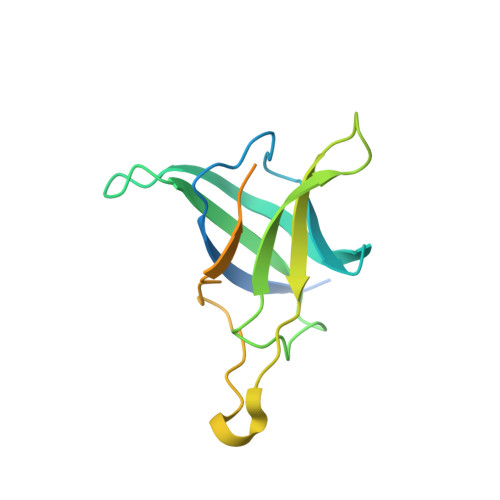
The membrane-associated solute-binding protein (SBP) MlaD of the maintenance of lipid asymmetry (Mla) system has been reported to help the transport of phospholipids (PLs) between the outer and inner membranes of Gram-negative bacteria. Despite the availability of structural information, the molecular mechanism underlying the transport of PLs and the ancestry of the protein MlaD remain unclear. In this study, we report the crystal structures of the periplasmic region of MlaD from Escherichia coli (EcMlaD) at a resolution range of 2.3-3.2 Å. The EcMlaD protomer consists of two distinct regions, viz. N-terminal β-barrel fold consisting of seven strands (referred to as MlaD domain) and C-terminal α-helical domain (HD). The protein EcMlaD oligomerizes to give rise to a homo-hexameric ring with a central channel that is hydrophobic and continuous with a variable diameter. Interestingly, the structural analysis revealed that the HD, instead of the MlaD domain, plays a critical role in determining the oligomeric state of the protein. Based on the analysis of available structural information, we propose a working mechanism of PL transport, viz. "asymmetric protomer movement (APM)". Wherein half of the EcMlaD hexamer would rise in the periplasmic side along with an outward movement of pore loops, resulting in the change of the central channel geometry. Furthermore, this study highlights that, unlike typical SBPs, EcMlaD possesses a fold similar to EF/AMT-type beta(6)-barrel and a unique ancestry. Altogether, the findings firmly establish EcMlaD to be a non-canonical SBP with a unique ligand-transport mechanism.
- Department of Biosciences and Bioengineering, Indian Institute of Technology Guwahati, Guwahati, Assam, 781039, India.
Organizational Affiliation: