8GXB

Crystal structure of NAD+ -II riboswitch in complex with NAD+


Experimental Data Snapshot

  • Method: X-RAY DIFFRACTION
  • Resolution: 2.15 Å
  • R-Value Free: 0.269 
  • R-Value Work: 0.219 
  • R-Value Observed: 0.222 

wwPDB Validation   3D Report Full Report


Ligand Structure Quality Assessment 


This is version 1.1 of the entry. See complete history


Literature

Structure-based investigations of the NAD+-II riboswitch.

Xu, X.Egger, M.Li, C.Chen, H.Micura, R.Ren, A.

(2023) Nucleic Acids Res 51: 54-67

  • DOI: https://doi.org/10.1093/nar/gkac1227
  • Primary Citation of Related Structures:  
    8GXB, 8GXC

  • PubMed Abstract: 

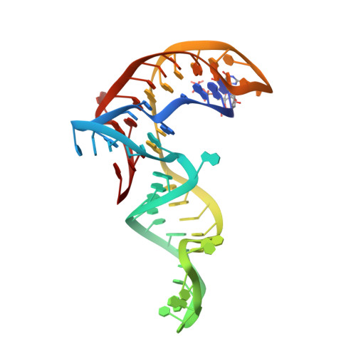
    Riboswitches are conserved non-coding domains in bacterial mRNA with gene regulation function that are essential for maintaining enzyme co-factor metabolism. Recently, the pnuC RNA motif was reported to selectively bind nicotinamide adenine dinucleotide (NAD+), defining a novel class of NAD+ riboswitches (NAD+-II) according to phylogenetic analysis. To reveal the three-dimensional architecture and the ligand-binding mode of this riboswitch, we solved the crystal structure of NAD+-II riboswitch in complex with NAD+. Strikingly and in contrast to class-I riboswitches that form a tight recognition pocket for the adenosine diphosphate (ADP) moiety of NAD+, the class-II riboswitches form a binding pocket for the nicotinamide mononucleotide (NMN) portion of NAD+ and display only unspecific interactions with the adenosine. We support this finding by an additional structure of the class-II RNA in complex with NMN alone. The structures define a novel RNA tertiary fold that was further confirmed by mutational analysis in combination with isothermal titration calorimetry (ITC), and 2-aminopurine-based fluorescence spectroscopic folding studies. Furthermore, we truncated the pnuC RNA motif to a short RNA helical scaffold with binding affinity comparable to the wild-type motif to allude to the potential of engineering the NAD+-II motif for biotechnological applications.


  • Organizational Affiliation

    Department of Gastroenterology/Department of Cardiology of the Second Affiliated Hospital, School of Medicine, Zhejiang University, Hangzhou, Zhejiang 310058, China.


Macromolecules

Find similar proteins by:  (by identity cutoff)  |  3D Structure
Entity ID: 2
MoleculeChains Sequence LengthOrganismDetailsImage
U1 small nuclear ribonucleoprotein A
C, D, E, F, G
97Homo sapiensMutation(s): 2 
Gene Names: SNRPA
UniProt & NIH Common Fund Data Resources
Find proteins for P09012 (Homo sapiens)
Explore P09012 
Go to UniProtKB:  P09012
PHAROS:  P09012
GTEx:  ENSG00000077312 
Entity Groups  
Sequence Clusters30% Identity50% Identity70% Identity90% Identity95% Identity100% Identity
UniProt GroupP09012
Sequence Annotations
Expand
  • Reference Sequence
Find similar nucleic acids by:  (by identity cutoff)  |  3D Structure
Entity ID: 1
MoleculeChains LengthOrganismImage
NAD+ II riboswitch
A, B
61Streptococcus sp.
Sequence Annotations
Expand
  • Reference Sequence
Small Molecules
Ligands 2 Unique
IDChains Name / Formula / InChI Key2D Diagram3D Interactions
NAD (Subject of Investigation/LOI)
Query on NAD

Download Ideal Coordinates CCD File 
H [auth A],
S [auth B]
NICOTINAMIDE-ADENINE-DINUCLEOTIDE
C21 H27 N7 O14 P2
BAWFJGJZGIEFAR-NNYOXOHSSA-N
MG
Query on MG

Download Ideal Coordinates CCD File 
AA [auth B]
BA [auth C]
I [auth A]
J [auth A]
K [auth A]
AA [auth B],
BA [auth C],
I [auth A],
J [auth A],
K [auth A],
L [auth A],
M [auth A],
N [auth A],
O [auth A],
P [auth A],
Q [auth A],
R [auth A],
T [auth B],
U [auth B],
V [auth B],
W [auth B],
X [auth B],
Y [auth B],
Z [auth B]
MAGNESIUM ION
Mg
JLVVSXFLKOJNIY-UHFFFAOYSA-N
Experimental Data & Validation

Experimental Data

  • Method: X-RAY DIFFRACTION
  • Resolution: 2.15 Å
  • R-Value Free: 0.269 
  • R-Value Work: 0.219 
  • R-Value Observed: 0.222 
  • Space Group: P 21 21 21
Unit Cell:
Length ( Å )Angle ( ˚ )
a = 57.46α = 90
b = 122.611β = 90
c = 154.649γ = 90
Software Package:
Software NamePurpose
PHENIXrefinement
HKL-2000data scaling
PDB_EXTRACTdata extraction
HKL-2000data reduction
PHASERphasing

Structure Validation

View Full Validation Report


Ligand Structure Quality Assessment 


Entry History & Funding Information

Deposition Data


Funding OrganizationLocationGrant Number
National Natural Science Foundation of China (NSFC)China--

Revision History  (Full details and data files)

  • Version 1.0: 2023-01-18
    Type: Initial release
  • Version 1.1: 2023-02-01
    Changes: Database references