8DBU | pdb_00008dbu

E. coli ATP synthase imaged in 10mM MgATP State2 "down" Fo classified


Experimental Data Snapshot

  • Method: ELECTRON MICROSCOPY
  • Resolution: 3.40 Å
  • Aggregation State: PARTICLE 
  • Reconstruction Method: SINGLE PARTICLE 

wwPDB Validation 3D Report Full Report

Validation slider image for 8DBU

This is version 1.2 of the entry. See complete history

Literature

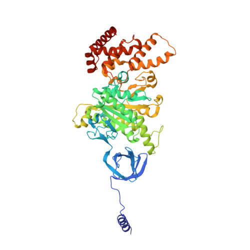
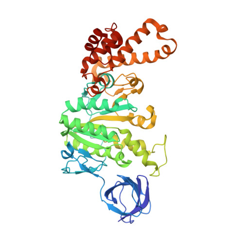
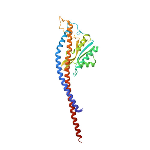
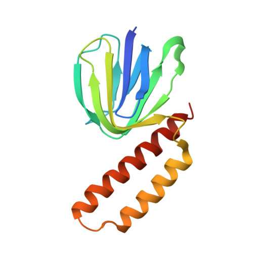
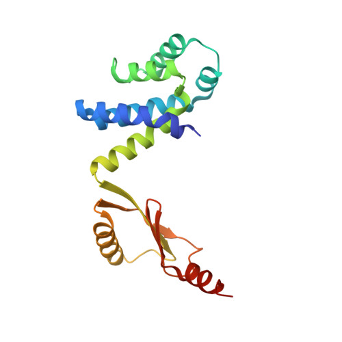
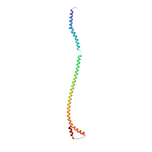
Changes within the central stalk of E. coli F 1 F o ATP synthase observed after addition of ATP.

Sobti, M.Zeng, Y.C.Walshe, J.L.Brown, S.H.J.Ishmukhametov, R.Stewart, A.G.

(2023) Commun Biol 6: 26-26

  • DOI: https://doi.org/10.1038/s42003-023-04414-z
  • Primary Citation Related Structures: 
    8DBP, 8DBQ, 8DBR, 8DBS, 8DBT, 8DBU, 8DBV, 8DBW

  • PubMed Abstract: 

    F 1 F o ATP synthase functions as a biological generator and makes a major contribution to cellular energy production. Proton flow generates rotation in the F o motor that is transferred to the F 1 motor to catalyze ATP production, with flexible F 1 /F o coupling required for efficient catalysis. F 1 F o ATP synthase can also operate in reverse, hydrolyzing ATP and pumping protons, and in bacteria this function can be regulated by an inhibitory ε subunit. Here we present cryo-EM data showing E. coli F 1 F o ATP synthase in different rotational and inhibited sub-states, observed following incubation with 10 mM MgATP. Our structures demonstrate how structural transitions within the inhibitory ε subunit induce torsional movement in the central stalk, thereby enabling its rotation within the F ο motor. This highlights the importance of the central rotor for flexible coupling of the F 1 and F o motors and provides further insight into the regulatory mechanism mediated by subunit ε.


  • Organizational Affiliation
    • Molecular, Structural and Computational Biology Division, The Victor Chang Cardiac Research Institute, Darlinghurst, NSW, Australia.

Macromolecule Content 

  • Total Structure Weight: 532.91 kDa 
  • Atom Count: 36,917 
  • Modeled Residue Count: 4,845 
  • Deposited Residue Count: 4,895 
  • Unique protein chains: 8

Macromolecules

Find similar proteins by:|  3D Structure
Entity ID: 1
MoleculeChains  Sequence LengthOrganismDetailsImage
ATP synthase subunit alpha
A, B, C
513Escherichia coliMutation(s): 0 
Gene Names: atpAECLG_02126
EC: 7.1.2.2
Membrane Entity: Yes 
UniProt
Find proteins for P0ABB0 (Escherichia coli (strain K12))
Explore P0ABB0 
Go to UniProtKB:  P0ABB0
Entity Groups
Sequence Clusters30% Identity50% Identity70% Identity90% Identity95% Identity100% Identity
UniProt GroupP0ABB0
Sequence Annotations
Expand
Reference Sequence
Find similar proteins by:|  3D Structure
Entity ID: 2
MoleculeChains  Sequence LengthOrganismDetailsImage
ATP synthase subunit beta
D, E, F
460Escherichia coliMutation(s): 0 
Gene Names: atpDEWT59_16525WLH_03015
EC: 7.1.2.2
Membrane Entity: Yes 
UniProt
Find proteins for P0ABB4 (Escherichia coli (strain K12))
Explore P0ABB4 
Go to UniProtKB:  P0ABB4
Entity Groups
Sequence Clusters30% Identity50% Identity70% Identity90% Identity95% Identity100% Identity
UniProt GroupP0ABB4
Sequence Annotations
Expand
Reference Sequence
Find similar proteins by:|  3D Structure
Entity ID: 3
MoleculeChains  Sequence LengthOrganismDetailsImage
ATP synthase gamma chain287Escherichia coliMutation(s): 0 
Membrane Entity: Yes 
UniProt
Find proteins for P0ABA6 (Escherichia coli (strain K12))
Explore P0ABA6 
Go to UniProtKB:  P0ABA6
Entity Groups
Sequence Clusters30% Identity50% Identity70% Identity90% Identity95% Identity100% Identity
UniProt GroupP0ABA6
Sequence Annotations
Expand
Reference Sequence
Find similar proteins by:|  3D Structure
Entity ID: 4
MoleculeChains  Sequence LengthOrganismDetailsImage
ATP synthase epsilon chain139Escherichia coliMutation(s): 0 
Gene Names: atpC
Membrane Entity: Yes 
UniProt
Find proteins for P0A6E6 (Escherichia coli (strain K12))
Explore P0A6E6 
Go to UniProtKB:  P0A6E6
Entity Groups
Sequence Clusters30% Identity50% Identity70% Identity90% Identity95% Identity100% Identity
UniProt GroupP0A6E6
Sequence Annotations
Expand
Reference Sequence
Find similar proteins by:|  3D Structure
Entity ID: 5
MoleculeChains  Sequence LengthOrganismDetailsImage
ATP synthase subunit c79Escherichia coliMutation(s): 0 
Gene Names: atpEECJG_03465
Membrane Entity: Yes 
UniProt
Find proteins for P68699 (Escherichia coli (strain K12))
Explore P68699 
Go to UniProtKB:  P68699
Entity Groups
Sequence Clusters30% Identity50% Identity70% Identity90% Identity95% Identity100% Identity
UniProt GroupP68699
Sequence Annotations
Expand
Reference Sequence
Find similar proteins by:|  3D Structure
Entity ID: 6
MoleculeChains  Sequence LengthOrganismDetailsImage
ATP synthase subunit deltaS [auth W]177Escherichia coliMutation(s): 0 
Gene Names: atpHHMPREF1611_00658
Membrane Entity: Yes 
UniProt
Find proteins for P0ABA4 (Escherichia coli (strain K12))
Explore P0ABA4 
Go to UniProtKB:  P0ABA4
Entity Groups
Sequence Clusters30% Identity50% Identity70% Identity90% Identity95% Identity100% Identity
UniProt GroupP0ABA4
Sequence Annotations
Expand
Reference Sequence
Find similar proteins by:|  3D Structure
Entity ID: 7
MoleculeChains  Sequence LengthOrganismDetailsImage
ATP synthase subunit bT [auth X],
U [auth Y]
156Escherichia coliMutation(s): 0 
Gene Names: atpf
Membrane Entity: Yes 
UniProt
Find proteins for P0ABA0 (Escherichia coli (strain K12))
Explore P0ABA0 
Go to UniProtKB:  P0ABA0
Entity Groups
Sequence Clusters30% Identity50% Identity70% Identity90% Identity95% Identity100% Identity
UniProt GroupP0ABA0
Sequence Annotations
Expand
Reference Sequence
Find similar proteins by:|  3D Structure
Entity ID: 8
MoleculeChains  Sequence LengthOrganismDetailsImage
ATP synthase subunit aV [auth a]271Escherichia coliMutation(s): 0 
Gene Names: atpB
Membrane Entity: Yes 
UniProt
Find proteins for P0AB98 (Escherichia coli (strain K12))
Explore P0AB98 
Go to UniProtKB:  P0AB98
Entity Groups
Sequence Clusters30% Identity50% Identity70% Identity90% Identity95% Identity100% Identity
UniProt GroupP0AB98
Sequence Annotations
Expand
Reference Sequence

Small Molecules

Ligands 3 Unique
IDChains Name / Formula / InChI Key2D Diagram3D Interactions
ATP

Query on ATP


Download:Ideal Coordinates CCD File
AA [auth C],
DA [auth D],
W [auth A],
Y [auth B]
ADENOSINE-5'-TRIPHOSPHATE
C10 H16 N5 O13 P3
ZKHQWZAMYRWXGA-KQYNXXCUSA-N
ADP

Query on ADP


Download:Ideal Coordinates CCD File
EA [auth E],
GA [auth F]
ADENOSINE-5'-DIPHOSPHATE
C10 H15 N5 O10 P2
XTWYTFMLZFPYCI-KQYNXXCUSA-N
MG

Query on MG


Download:Ideal Coordinates CCD File
BA [auth C],
CA [auth D],
FA [auth E],
X [auth A],
Z [auth B]
MAGNESIUM ION
Mg
JLVVSXFLKOJNIY-UHFFFAOYSA-N

Experimental Data & Validation

Experimental Data

  • Method: ELECTRON MICROSCOPY
  • Resolution: 3.40 Å
  • Aggregation State: PARTICLE 
  • Reconstruction Method: SINGLE PARTICLE 
EM Software:
TaskSoftware PackageVersion
MODEL REFINEMENTPHENIX

Structure Validation

View Full Validation Report


Entry History 

& Funding Information

Deposition Data


Funding OrganizationLocationGrant Number
National Health and Medical Research Council (NHMRC, Australia)Australia--

Revision History  (Full details and data files)

  • Version 1.0: 2023-01-25
    Type: Initial release
  • Version 1.1: 2024-06-12
    Changes: Data collection
  • Version 1.2: 2025-05-21
    Changes: Data collection, Structure summary