8CS9

Composite reconstruction of Class 1 of the erythrocyte ankyrin-1 complex


Experimental Data Snapshot

  • Method: ELECTRON MICROSCOPY
  • Resolution: 2.74 Å
  • Aggregation State: PARTICLE 
  • Reconstruction Method: SINGLE PARTICLE 

wwPDB Validation   3D Report Full Report


This is version 1.1 of the entry. See complete history


Literature

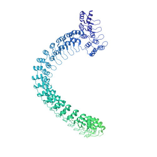
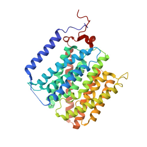
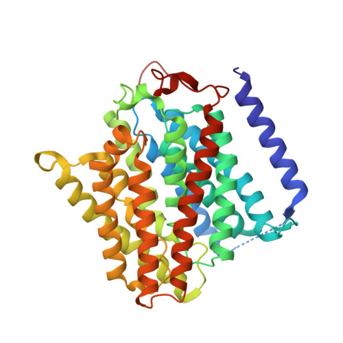
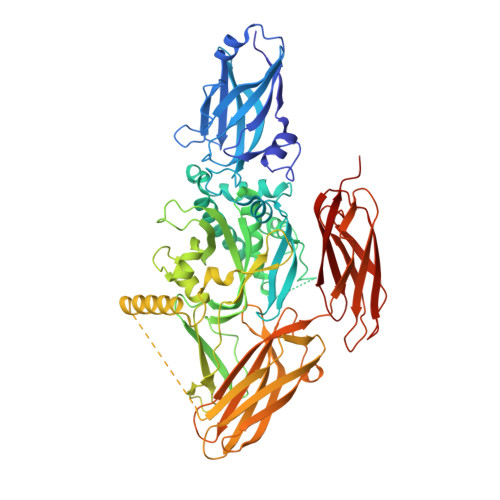
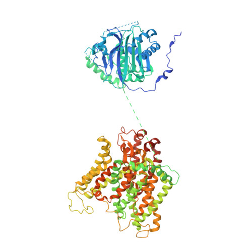
Architecture of the human erythrocyte ankyrin-1 complex.

Vallese, F.Kim, K.Yen, L.Y.Johnston, J.D.Noble, A.J.Cali, T.Clarke, O.B.

(2022) Nat Struct Mol Biol 29: 706-718

  • DOI: https://doi.org/10.1038/s41594-022-00792-w
  • Primary Citation of Related Structures:  
    7UZ3, 7UZE, 7UZQ, 7UZS, 7UZU, 7UZV, 7V07, 7V0K, 7V0M, 7V0Q, 7V0S, 7V0T, 7V0U, 7V0X, 7V0Y, 7V19, 8CRQ, 8CRR, 8CRT, 8CS9, 8CSL, 8CSV, 8CSW, 8CSX, 8CSY, 8CT2, 8CT3, 8CTE

  • PubMed Abstract: 

    The stability and shape of the erythrocyte membrane is provided by the ankyrin-1 complex, but how it tethers the spectrin-actin cytoskeleton to the lipid bilayer and the nature of its association with the band 3 anion exchanger and the Rhesus glycoproteins remains unknown. Here we present structures of ankyrin-1 complexes purified from human erythrocytes. We reveal the architecture of a core complex of ankyrin-1, the Rhesus proteins RhAG and RhCE, the band 3 anion exchanger, protein 4.2, glycophorin A and glycophorin B. The distinct T-shaped conformation of membrane-bound ankyrin-1 facilitates recognition of RhCE and, unexpectedly, the water channel aquaporin-1. Together, our results uncover the molecular details of ankyrin-1 association with the erythrocyte membrane, and illustrate the mechanism of ankyrin-mediated membrane protein clustering.


  • Organizational Affiliation

    Department of Anesthesiology, Columbia University Irving Medical Center, New York, NY, USA.


Macromolecules
Find similar proteins by:  (by identity cutoff)  |  3D Structure
Entity ID: 1
MoleculeChains Sequence LengthOrganismDetailsImage
Ankyrin-11,881Homo sapiensMutation(s): 0 
Membrane Entity: Yes 
UniProt & NIH Common Fund Data Resources
Find proteins for P16157 (Homo sapiens)
Explore P16157 
Go to UniProtKB:  P16157
PHAROS:  P16157
GTEx:  ENSG00000029534 
Entity Groups  
Sequence Clusters30% Identity50% Identity70% Identity90% Identity95% Identity100% Identity
UniProt GroupP16157
Sequence Annotations
Expand
  • Reference Sequence
Find similar proteins by:  (by identity cutoff)  |  3D Structure
Entity ID: 2
MoleculeChains Sequence LengthOrganismDetailsImage
Blood group Rh(CE) polypeptideB [auth K]417Homo sapiensMutation(s): 0 
Membrane Entity: Yes 
UniProt & NIH Common Fund Data Resources
Find proteins for P18577 (Homo sapiens)
Explore P18577 
Go to UniProtKB:  P18577
PHAROS:  P18577
GTEx:  ENSG00000188672 
Entity Groups  
Sequence Clusters30% Identity50% Identity70% Identity90% Identity95% Identity100% Identity
UniProt GroupP18577
Sequence Annotations
Expand
  • Reference Sequence
Find similar proteins by:  (by identity cutoff)  |  3D Structure
Entity ID: 3
MoleculeChains Sequence LengthOrganismDetailsImage
Ammonium transporter Rh type AC [auth L],
D [auth Q]
409Homo sapiensMutation(s): 0 
Membrane Entity: Yes 
UniProt & NIH Common Fund Data Resources
Find proteins for Q02094 (Homo sapiens)
Explore Q02094 
Go to UniProtKB:  Q02094
PHAROS:  Q02094
GTEx:  ENSG00000112077 
Entity Groups  
Sequence Clusters30% Identity50% Identity70% Identity90% Identity95% Identity100% Identity
UniProt GroupQ02094
Sequence Annotations
Expand
  • Reference Sequence
Find similar proteins by:  (by identity cutoff)  |  3D Structure
Entity ID: 4
MoleculeChains Sequence LengthOrganismDetailsImage
Protein 4.2E [auth X]691Homo sapiensMutation(s): 0 
Membrane Entity: Yes 
UniProt & NIH Common Fund Data Resources
Find proteins for P16452 (Homo sapiens)
Explore P16452 
Go to UniProtKB:  P16452
PHAROS:  P16452
GTEx:  ENSG00000166947 
Entity Groups  
Sequence Clusters30% Identity50% Identity70% Identity90% Identity95% Identity100% Identity
UniProt GroupP16452
Sequence Annotations
Expand
  • Reference Sequence
Find similar proteins by:  (by identity cutoff)  |  3D Structure
Entity ID: 5
MoleculeChains Sequence LengthOrganismDetailsImage
Glycophorin-BF [auth P]91Homo sapiensMutation(s): 0 
Membrane Entity: Yes 
UniProt & NIH Common Fund Data Resources
Find proteins for P06028 (Homo sapiens)
Explore P06028 
Go to UniProtKB:  P06028
PHAROS:  P06028
GTEx:  ENSG00000250361 
Entity Groups  
Sequence Clusters30% Identity50% Identity70% Identity90% Identity95% Identity100% Identity
UniProt GroupP06028
Sequence Annotations
Expand
  • Reference Sequence
Find similar proteins by:  (by identity cutoff)  |  3D Structure
Entity ID: 6
MoleculeChains Sequence LengthOrganismDetailsImage
Glycophorin-A150Homo sapiensMutation(s): 0 
Membrane Entity: Yes 
UniProt & NIH Common Fund Data Resources
Find proteins for P02724 (Homo sapiens)
Explore P02724 
Go to UniProtKB:  P02724
PHAROS:  P02724
GTEx:  ENSG00000170180 
Entity Groups  
Sequence Clusters30% Identity50% Identity70% Identity90% Identity95% Identity100% Identity
UniProt GroupP02724
Sequence Annotations
Expand
  • Reference Sequence
Find similar proteins by:  (by identity cutoff)  |  3D Structure
Entity ID: 7
MoleculeChains Sequence LengthOrganismDetailsImage
Band 3 anion transport protein911Homo sapiensMutation(s): 0 
Membrane Entity: Yes 
UniProt & NIH Common Fund Data Resources
Find proteins for P02730 (Homo sapiens)
Explore P02730 
Go to UniProtKB:  P02730
PHAROS:  P02730
GTEx:  ENSG00000004939 
Entity Groups  
Sequence Clusters30% Identity50% Identity70% Identity90% Identity95% Identity100% Identity
UniProt GroupP02730
Sequence Annotations
Expand
  • Reference Sequence
Oligosaccharides

Help

Entity ID: 8
MoleculeChains Length2D Diagram Glycosylation3D Interactions
2-acetamido-2-deoxy-beta-D-glucopyranose-(1-4)-2-acetamido-2-deoxy-beta-D-glucopyranoseS [auth B],
T [auth C],
U [auth D]
2N/AN-Glycosylation
Glycosylation Resources
GlyTouCan:  G42666HT
GlyCosmos:  G42666HT
GlyGen:  G42666HT
Small Molecules
Ligands 4 Unique
IDChains Name / Formula / InChI Key2D Diagram3D Interactions
AJP
Query on AJP

Download Ideal Coordinates CCD File 
X [auth Q]Digitonin
C56 H92 O29
UVYVLBIGDKGWPX-KUAJCENISA-N
PIO (Subject of Investigation/LOI)
Query on PIO

Download Ideal Coordinates CCD File 
BA [auth V]
DA [auth e]
HA [auth Y]
JA [auth f]
OA [auth g]
BA [auth V],
DA [auth e],
HA [auth Y],
JA [auth f],
OA [auth g],
PA [auth g]
[(2R)-2-octanoyloxy-3-[oxidanyl-[(1R,2R,3S,4R,5R,6S)-2,3,6-tris(oxidanyl)-4,5-diphosphonooxy-cyclohexyl]oxy-phosphoryl]oxy-propyl] octanoate
C25 H49 O19 P3
XLNCEHRXXWQMPK-MJUMVPIBSA-N
CLR
Query on CLR

Download Ideal Coordinates CCD File 
CA [auth e]
EA [auth Y]
IA [auth f]
KA [auth c]
LA [auth Z]
CA [auth e],
EA [auth Y],
IA [auth f],
KA [auth c],
LA [auth Z],
V [auth L],
W [auth L],
Y [auth V]
CHOLESTEROL
C27 H46 O
HVYWMOMLDIMFJA-DPAQBDIFSA-N
NAG
Query on NAG

Download Ideal Coordinates CCD File 
AA [auth V]
FA [auth Y]
GA [auth Y]
MA [auth Z]
NA [auth Z]
AA [auth V],
FA [auth Y],
GA [auth Y],
MA [auth Z],
NA [auth Z],
Z [auth V]
2-acetamido-2-deoxy-beta-D-glucopyranose
C8 H15 N O6
OVRNDRQMDRJTHS-FMDGEEDCSA-N
Experimental Data & Validation

Experimental Data

  • Method: ELECTRON MICROSCOPY
  • Resolution: 2.74 Å
  • Aggregation State: PARTICLE 
  • Reconstruction Method: SINGLE PARTICLE 
EM Software:
TaskSoftware PackageVersion
RECONSTRUCTIONcryoSPARC

Structure Validation

View Full Validation Report


Entry History & Funding Information

Deposition Data


Funding OrganizationLocationGrant Number
Not funded--

Revision History  (Full details and data files)

  • Version 1.0: 2022-07-20
    Type: Initial release
  • Version 1.1: 2022-07-27
    Changes: Database references