8A65 | pdb_00008a65

Small molecule stabilizer (compound 3) for FOXO1 and 14-3-3


Experimental Data Snapshot

  • Method: X-RAY DIFFRACTION
  • Resolution: 1.60 Å
  • R-Value Free: 
    0.193 (Depositor), 0.216 (DCC) 
  • R-Value Work: 
    0.167 (Depositor) 
  • R-Value Observed: 
    0.168 (Depositor) 

Starting Model: experimental
View more details

wwPDB Validation 3D Report Full Report

Validation slider image for 8A65

Ligand Structure Quality Assessment 


This is version 1.3 of the entry. See complete history

Literature

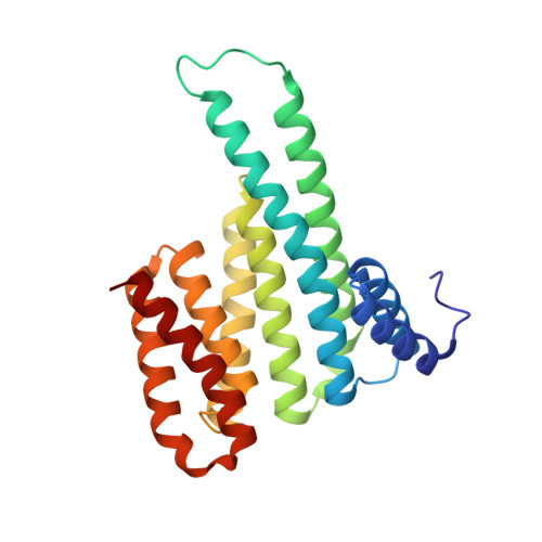
A Systematic Approach to the Discovery of Protein-Protein Interaction Stabilizers.

Kenanova, D.N.Visser, E.J.Virta, J.M.Sijbesma, E.Centorrino, F.Vickery, H.R.Zhong, M.Neitz, R.J.Brunsveld, L.Ottmann, C.Arkin, M.R.

(2023) ACS Cent Sci 9: 937-946

  • DOI: https://doi.org/10.1021/acscentsci.2c01449
  • Primary Citation Related Structures: 
    8A62, 8A65, 8A68, 8A6F, 8A6H, 8ADM, 8AFN, 8AV0

  • PubMed Abstract: 

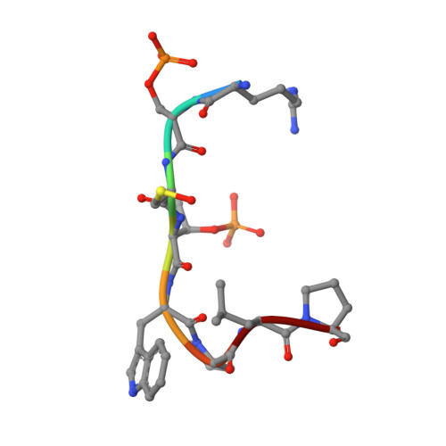
    Dysregulation of protein-protein interactions (PPIs) commonly leads to disease. PPI stabilization has only recently been systematically explored for drug discovery despite being a powerful approach to selectively target intrinsically disordered proteins and hub proteins, like 14-3-3, with multiple interaction partners. Disulfide tethering is a site-directed fragment-based drug discovery (FBDD) methodology for identifying reversibly covalent small molecules. We explored the scope of disulfide tethering for the discovery of selective PPI stabilizers (molecular glues) using the hub protein 14-3-3σ. We screened complexes of 14-3-3 with 5 biologically and structurally diverse phosphopeptides derived from the 14-3-3 client proteins ERα, FOXO1, C-RAF, USP8, and SOS1. Stabilizing fragments were found for 4/5 client complexes. Structural elucidation of these complexes revealed the ability of some peptides to conformationally adapt to make productive interactions with the tethered fragments. We validated eight fragment stabilizers, six of which showed selectivity for one phosphopeptide client, and structurally characterized two nonselective hits and four fragments that selectively stabilized C-RAF or FOXO1. The most efficacious fragment increased 14-3-3σ/C-RAF phosphopeptide affinity by 430-fold. Disulfide tethering to the wildtype C38 in 14-3-3σ provided diverse structures for future optimization of 14-3-3/client stabilizers and highlighted a systematic method to discover molecular glues.


  • Organizational Affiliation
    • Department of Pharmaceutical Chemistry and Small Molecule Discovery Center (SMDC), University of California, San Francisco 94143, United States.

Macromolecule Content 

  • Total Structure Weight: 28.21 kDa 
  • Atom Count: 2,269 
  • Modeled Residue Count: 244 
  • Deposited Residue Count: 244 
  • Unique protein chains: 2

Macromolecules

Find similar proteins by:|  3D Structure
Entity ID: 1
MoleculeChains  Sequence LengthOrganismDetailsImage
14-3-3 protein sigma236Homo sapiensMutation(s): 0 
Gene Names: SFNHME1
UniProt & NIH Common Fund Data Resources
Find proteins for P31947 (Homo sapiens)
Explore P31947 
Go to UniProtKB:  P31947
PHAROS:  P31947
GTEx:  ENSG00000175793 
Entity Groups
Sequence Clusters30% Identity50% Identity70% Identity90% Identity95% Identity100% Identity
UniProt GroupP31947
Sequence Annotations
Expand
Reference Sequence
Find similar proteins by:  Sequence   |   3D Structure  
Entity ID: 2
MoleculeChains  Sequence LengthOrganismDetailsImage
Forkhead box protein O18Homo sapiensMutation(s): 0 
UniProt & NIH Common Fund Data Resources
Find proteins for Q12778 (Homo sapiens)
Explore Q12778 
Go to UniProtKB:  Q12778
PHAROS:  Q12778
GTEx:  ENSG00000150907 
Entity Groups
UniProt GroupQ12778
Sequence Annotations
Expand
Reference Sequence

Small Molecules

Modified Residues  3 Unique
IDChains TypeFormula2D DiagramParent
CSO
Query on CSO
B
L-PEPTIDE LINKINGC3 H7 N O3 SCYS
SEP
Query on SEP
B
L-PEPTIDE LINKINGC3 H8 N O6 PSER
TPO
Query on TPO
B
L-PEPTIDE LINKINGC4 H10 N O6 PTHR

Experimental Data & Validation

Experimental Data

  • Method: X-RAY DIFFRACTION
  • Resolution: 1.60 Å
  • R-Value Free:  0.193 (Depositor), 0.216 (DCC) 
  • R-Value Work:  0.167 (Depositor) 
  • R-Value Observed: 0.168 (Depositor) 
Space Group: C 2 2 21
Unit Cell:
Length ( Å )Angle ( ˚ )
a = 81.879α = 90
b = 112.144β = 90
c = 62.632γ = 90
Software Package:
Software NamePurpose
REFMACrefinement
PDB_EXTRACTdata extraction
HKL-2000data reduction
Aimlessdata scaling
MOLREPphasing

Structure Validation

View Full Validation Report


Ligand Structure Quality Assessment 


Entry History 

& Funding Information

Deposition Data


Funding OrganizationLocationGrant Number
Netherlands Organisation for Scientific Research (NWO)Netherlands024.001.035

Revision History  (Full details and data files)

  • Version 1.0: 2023-04-26
    Type: Initial release
  • Version 1.1: 2023-06-07
    Changes: Database references
  • Version 1.2: 2024-02-07
    Changes: Data collection, Refinement description
  • Version 1.3: 2024-11-13
    Changes: Structure summary