8U22 | pdb_00008u22

A Highly Stable Variant of Corynactis Californica Green Fluorescent Protein, ccGFP 7


Experimental Data Snapshot

  • Method: X-RAY DIFFRACTION
  • Resolution: 1.80 Å
  • R-Value Free: 
    0.210 (Depositor), 0.210 (DCC) 
  • R-Value Work: 
    0.180 (Depositor), 0.182 (DCC) 
  • R-Value Observed: 
    0.183 (Depositor) 

Starting Model: experimental
View more details

wwPDB Validation 3D Report Full Report

Validation slider image for 8U22

This is version 3.0 of the entry. See complete history

Literature

Engineering highly stable variants of Corynactis californica green fluorescent proteins.

Hung, L.W.Terwilliger, T.C.Waldo, G.S.Nguyen, H.B.

(2024) Protein Sci 33: e4886-e4886

  • DOI: https://doi.org/10.1002/pro.4886
  • Primary Citation Related Structures: 
    8U20, 8U21, 8U22, 8U23, 8U24

  • PubMed Abstract: 

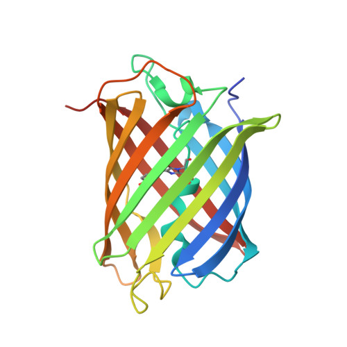
    Fluorescent proteins (FPs) are versatile biomarkers that facilitate effective detection and tracking of macromolecules of interest in real time. Engineered FPs such as superfolder green fluorescent protein (sfGFP) and superfolder Cherry (sfCherry) have exceptional refolding capability capable of delivering fluorescent readout in harsh environments where most proteins lose their native functions. Our recent work on the development of a split FP from a species of strawberry anemone, Corynactis californica, delivered pairs of fragments with up to threefold faster complementation than split GFP. We present the biophysical, biochemical, and structural characteristics of five full-length variants derived from these split C. californica GFP (ccGFP). These ccGFP variants are more tolerant under chemical denaturation with up to 8 kcal/mol lower unfolding free energy than that of the sfGFP. It is likely that some of these ccGFP variants could be suitable as biomarkers under more adverse environments where sfGFP fails to survive. A structural analysis suggests explanations of the variations in stabilities among the ccGFP variants.


  • Organizational Affiliation
    • Bioscience Division, MS M888, Los Alamos National Laboratory, Los Alamos, New Mexico, USA.

Macromolecule Content 

  • Total Structure Weight: 25.04 kDa 
  • Atom Count: 1,993 
  • Modeled Residue Count: 212 
  • Deposited Residue Count: 219 
  • Unique protein chains: 1

Macromolecules

Find similar proteins by:|  3D Structure
Entity ID: 1
MoleculeChains  Sequence LengthOrganismDetailsImage
Green Fluorescent Protein Variant #7, ccGFP 7219Corynactis californicaMutation(s): 0 
UniProt
Find proteins for Q1ALD4 (Corynactis californica)
Explore Q1ALD4 
Go to UniProtKB:  Q1ALD4
Entity Groups
Sequence Clusters30% Identity50% Identity70% Identity90% Identity95% Identity100% Identity
UniProt GroupQ1ALD4
Sequence Annotations
Expand
Reference Sequence

Small Molecules

Modified Residues  1 Unique
IDChains TypeFormula2D DiagramParent
C12
Query on C12
A
L-PEPTIDE LINKINGC15 H18 N3 O5THR, TYR, GLY

Experimental Data & Validation

Experimental Data

  • Method: X-RAY DIFFRACTION
  • Resolution: 1.80 Å
  • R-Value Free:  0.210 (Depositor), 0.210 (DCC) 
  • R-Value Work:  0.180 (Depositor), 0.182 (DCC) 
  • R-Value Observed: 0.183 (Depositor) 
Space Group: P 43 21 2
Unit Cell:
Length ( Å )Angle ( ˚ )
a = 54.582α = 90
b = 54.582β = 90
c = 169.303γ = 90
Software Package:
Software NamePurpose
PHENIXrefinement
HKL-2000data scaling
HKL-2000data reduction
PHASERphasing

Structure Validation

View Full Validation Report


Entry History 

& Funding Information

Deposition Data


Funding OrganizationLocationGrant Number
National Institutes of Health/National Institute of General Medical Sciences (NIH/NIGMS)United States--

Revision History  (Full details and data files)

  • Version 1.0: 2024-01-31
    Type: Initial release
  • Version 1.1: 2024-02-07
    Changes: Database references
  • Version 2.0: 2024-07-10
    Changes: Data collection, Derived calculations, Non-polymer description, Structure summary
  • Version 2.1: 2024-10-23
    Changes: Structure summary
  • Version 3.0: 2026-03-18
    Changes: Polymer sequence