8TN9

Structural architecture of the acidic region of the B domain of coagulation factor V

  • Classification: BLOOD CLOTTING
  • Organism(s): Homo sapiens
  • Mutation(s): No 

  • Deposited: 2023-08-01 Released: 2023-10-04 
  • Deposition Author(s): Mohammed, B.M., Basore, K., Summers, B., Pelc, L.A., Di Cera, E.
  • Funding Organization(s): National Institutes of Health/National Heart, Lung, and Blood Institute (NIH/NHLBI), Childrens Discovery Institute of Washington University and St. Louis Childrens Hospital, The Foundation for Barnes-Jewish Hospital, National Institutes of Health/National Institute of Diabetes and Digestive and Kidney Disease (NIH/NIDDK), National Institutes of Health/National Cancer Institute (NIH/NCI)

Experimental Data Snapshot

  • Method: ELECTRON MICROSCOPY
  • Resolution: 3.05 Å
  • Aggregation State: PARTICLE 
  • Reconstruction Method: SINGLE PARTICLE 

wwPDB Validation   3D Report Full Report


This is version 1.2 of the entry. See complete history


Literature

Structural architecture of the acidic region of the B domain of coagulation factor V.

Mohammed, B.M.Basore, K.Summers, B.Pelc, L.A.Di Cera, E.

(2024) J Thromb Haemost 22: 709-714

  • DOI: https://doi.org/10.1016/j.jtha.2023.11.003
  • Primary Citation of Related Structures:  
    8TN9

  • PubMed Abstract: 

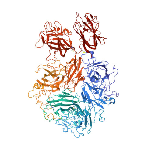
    Coagulation factor (F)V features an A1-A2-B-A3-C1-C2 domain organization and functions as the inactive precursor of FVa, a component of the prothrombinase complex required for rapid thrombin generation in the penultimate step of the coagulation cascade. An intramolecular interaction within the large B domain (residues 710-1545) involves the basic region (BR, residues 963-1008) and acidic region (AR, residues 1493-1537) and locks FV in its inactive state. However, structural information on this important regulatory interaction or on the separate architecture of the AR and BR remains elusive due to conformational disorder of the B domain.


  • Organizational Affiliation

    Edward A. Doisy Department of Biochemistry and Molecular Biology, Saint Louis University School of Medicine, St. Louis, Missouri, USA.


Macromolecules
Find similar proteins by:  (by identity cutoff)  |  3D Structure
Entity ID: 1
MoleculeChains Sequence LengthOrganismDetailsImage
Coagulation factor V2,196Homo sapiensMutation(s): 0 
UniProt & NIH Common Fund Data Resources
Find proteins for P12259 (Homo sapiens)
Explore P12259 
Go to UniProtKB:  P12259
PHAROS:  P12259
GTEx:  ENSG00000198734 
Entity Groups  
Sequence Clusters30% Identity50% Identity70% Identity90% Identity95% Identity100% Identity
UniProt GroupP12259
Sequence Annotations
Expand
  • Reference Sequence
Experimental Data & Validation

Experimental Data

  • Method: ELECTRON MICROSCOPY
  • Resolution: 3.05 Å
  • Aggregation State: PARTICLE 
  • Reconstruction Method: SINGLE PARTICLE 
EM Software:
TaskSoftware PackageVersion
RECONSTRUCTIONcryoSPARC4.2.1
MODEL REFINEMENTcryoSPARC4.2.1
MODEL REFINEMENTPHENIX1.20.1

Structure Validation

View Full Validation Report


Entry History & Funding Information

Deposition Data


Funding OrganizationLocationGrant Number
National Institutes of Health/National Heart, Lung, and Blood Institute (NIH/NHLBI)United StatesHL049413, HL139554 and HL147821
Childrens Discovery Institute of Washington University and St. Louis Childrens HospitalUnited StatesCDI-CORE-2015-505 and CDI-CORE-2019-813
The Foundation for Barnes-Jewish HospitalUnited States3770
National Institutes of Health/National Institute of Diabetes and Digestive and Kidney Disease (NIH/NIDDK)United StatesDK020579
National Institutes of Health/National Cancer Institute (NIH/NCI)United StatesCA091842

Revision History  (Full details and data files)

  • Version 1.0: 2023-10-04
    Type: Initial release
  • Version 1.1: 2023-12-20
    Changes: Database references
  • Version 1.2: 2024-03-13
    Changes: Database references