Structural Basis of PML-RARA Oncoprotein Targeting by Arsenic Unravels a Cysteine Rheostat Controlling PML Body Assembly and Function.
Bercier, P., Wang, Q.Q., Zang, N., Zhang, J., Yang, C., Maimaitiyiming, Y., Abou-Ghali, M., Berthier, C., Wu, C., Niwa-Kawakita, M., Dirami, T., Geoffroy, M.C., Ferhi, O., Quentin, S., Benhenda, S., Ogra, Y., Gueroui, Z., Zhou, C., Naranmandura, H., de The, H., Lallemand-Breitenbach, V.(2023) Cancer Discov 13: 2548-2565
- PubMed: 37655965 Search on PubMedSearch on PubMed Central
- DOI: https://doi.org/10.1158/2159-8290.CD-23-0453
- Primary Citation Related Structures:
8J25, 8J2P - PubMed Abstract:
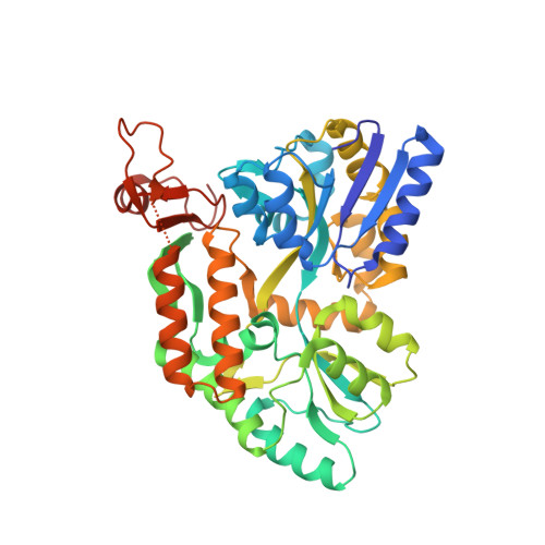
PML nuclear bodies (NB) are disrupted in PML-RARA-driven acute promyelocytic leukemia (APL). Arsenic trioxide (ATO) cures 70% of patients with APL, driving PML-RARA degradation and NB reformation. In non-APL cells, arsenic binding onto PML also amplifies NB formation. Yet, the actual molecular mechanism(s) involved remain(s) elusive. Here, we establish that PML NBs display some features of liquid-liquid phase separation and that ATO induces a gel-like transition. PML B-box-2 structure reveals an alpha helix driving B2 trimerization and positioning a cysteine trio to form an ideal arsenic-binding pocket. Altering either of the latter impedes ATO-driven NB assembly, PML sumoylation, and PML-RARA degradation, mechanistically explaining clinical ATO resistance. This B2 trimer and the C213 trio create an oxidation-sensitive rheostat that controls PML NB assembly dynamics and downstream signaling in both basal state and during stress response. These findings identify the structural basis for arsenic targeting of PML that could pave the way to novel cancer drugs. Arsenic curative effects in APL rely on PML targeting. We report a PML B-box-2 structure that drives trimer assembly, positioning a cysteine trio to form an arsenic-binding pocket, which is disrupted in resistant patients. Identification of this ROS-sensitive triad controlling PML dynamics and functions could yield novel drugs. See related commentary by Salomoni, p. 2505. This article is featured in Selected Articles from This Issue, p. 2489.
- Center for Interdisciplinary Research in Biology (CIRB), Collège de France, CNRS, INSERM, Université PSL, Paris, France.
Organizational Affiliation: