Mutational and structural analyses of UdgX: insights into the active site pocket architecture and its evolution.
Aroli, S., Woo, E.J., Gopal, B., Varshney, U.(2023) Nucleic Acids Res 51: 6554-6565
- PubMed: 37283083 Search on PubMedSearch on PubMed Central
- DOI: https://doi.org/10.1093/nar/gkad486
- Primary Citation Related Structures:
8IIE, 8IIF, 8IIG, 8IIH, 8III, 8IIJ, 8IIL, 8IIM, 8IIN, 8IIO, 8IIP, 8IIQ, 8IIR, 8IIS, 8IIT - PubMed Abstract:
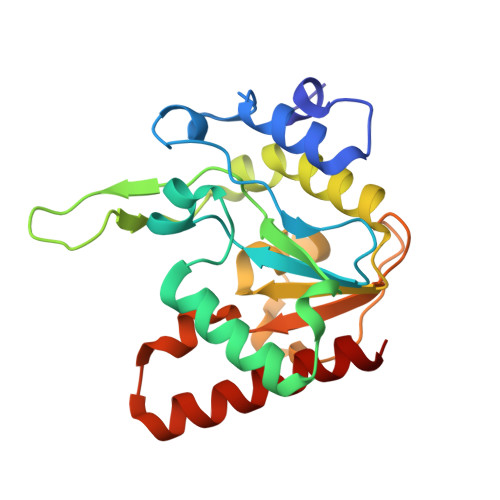
UdgX excises uracil from uracil-containing DNA to concurrently form a covalent bond with the resulting AP-DNA. Structurally, UdgX is highly similar to family-4 UDGs (F4-UDGs). However, UdgX is unique in possessing a flexible R-loop (105KRRIH109). Among the class-defining motifs, while its motif A (51GEQPG55) diverged to possess Q53 in place of A53/G53 in F4-UDGs, motif B [178HPS(S/A)(L/V)(L/V)R184] has remained unchanged. Previously, we proposed an SN1 mechanism resulting in a covalent bond between H109 and AP-DNA. In this study, we investigated several single/double mutants of UdgX. The H109A, H109S, H109G, H109Q, H109C and H109K mutants gain conventional UDG activity to varying levels. The crystal structures of UdgX mutants show topological changes in their active sites, rationalizing their UDG activities. The E52Q, E52N and E52A mutants reveal that E52 forms a catalytic dyad with H109 to enhance its nucleophilicity. The Q53A mutant supports that UdgX specific evolution of Q53 occurred essentially to stabilize the R-loop conformation. The R184A mutation (motif B) supports the role of R184 in substrate-binding. Taken together, the structural, bioinformatics, and mutational studies suggest that UdgX diverged from F4-UDGs, and the emergence of the characteristic R-loop in UdgX is functionally assisted by A53/G53 to Q53 changes in motif A.
- Department of Microbiology and Cell Biology, Indian Institute of Science, Bangalore 560012, India.
Organizational Affiliation: