Red fluorescent proteins engineered from green fluorescent proteins.
Imamura, H., Otsubo, S., Nishida, M., Takekawa, N., Imada, K.(2023) Proc Natl Acad Sci U S A 120: e2307687120-e2307687120
- PubMed: 37871160 Search on PubMedSearch on PubMed Central
- DOI: https://doi.org/10.1073/pnas.2307687120
- Primary Citation Related Structures:
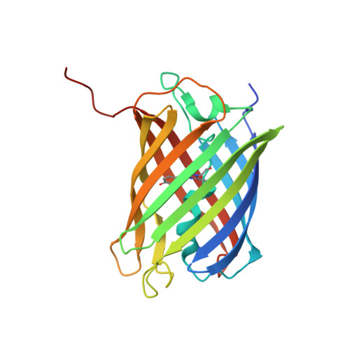
8I4J, 8I4K - PubMed Abstract:
Fluorescent proteins (FPs) form a fluorophore through autocatalysis from three consecutive amino acid residues within a polypeptide chain. The two major groups, green FPs (GFPs) and red FPs (RFPs), have distinct fluorophore structures; RFPs have an extended π-conjugation system with an additional double bond. However, due to the low sequence homology between the two groups, amino acid residues essential for determining the different fluorophore structures were unclear. Therefore, engineering a GFP into an RFP has been challenging, and the exact mechanism of how GFPs and RFPs achieve different autocatalytic reactions remained elucidated. Here, we show the conversion of two coral GFPs, AzamiGreen (AG) and mcavGFP, into RFPs by defined mutations. Structural comparison of AG and AzamiRed1.0, an AG-derived RFP, revealed that the mutations triggered drastic rearrangements in the interaction networks between amino acid residues around the fluorophore, suggesting that coordinated multisite mutations are required for the green-to-red conversion. As a result of the structural rearrangements, a cavity suitable for the entry of an oxygen molecule, which is necessary for the double bond formation of the red fluorophores, is created in the proximity of the fluorophore. We also show that a monomeric variant of AzamiRed1.0 can be used for labeling organelles and proteins in mammalian cells. Our results provide a structural basis for understanding the red fluorophore formation mechanism and demonstrate that protein engineering of GFPs is a promising way to create RFPs suitable for fluorescent tags.
- Division of Systemic Life Science, Graduate School of Biostudies, Kyoto University, Kyoto 606-8501, Japan.
Organizational Affiliation: