8HHA | pdb_00008hha

F1 domain of FoF1-ATPase from Bacillus PS3,120 degrees,lowATP


Experimental Data Snapshot

  • Method: ELECTRON MICROSCOPY
  • Resolution: 3.40 Å
  • Aggregation State: PARTICLE 
  • Reconstruction Method: SINGLE PARTICLE 

Starting Model: experimental
View more details

wwPDB Validation   3D Report Full Report


This is version 1.2 of the entry. See complete history


Literature

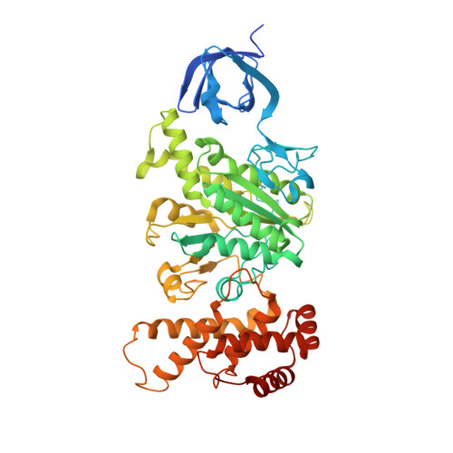
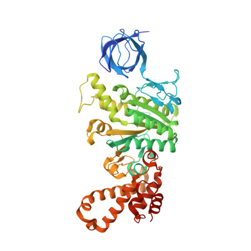
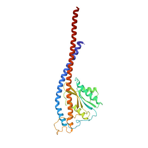
Mechanism of ATP hydrolysis dependent rotation of bacterial ATP synthase.

Nakano, A.Kishikawa, J.I.Mitsuoka, K.Yokoyama, K.

(2023) Nat Commun 14: 4090-4090

  • DOI: https://doi.org/10.1038/s41467-023-39742-5
  • Primary Citation of Related Structures:  
    8HH1, 8HH2, 8HH3, 8HH4, 8HH5, 8HH6, 8HH7, 8HH8, 8HH9, 8HHA, 8HHB, 8HHC

  • PubMed Abstract: 

    F 1 domain of ATP synthase is a rotary ATPase complex in which rotation of central γ-subunit proceeds in 120° steps against a surrounding α 3 β 3 fueled by ATP hydrolysis. How the ATP hydrolysis reactions occurring in three catalytic αβ dimers are coupled to mechanical rotation is a key outstanding question. Here we describe catalytic intermediates of the F 1 domain in F o F 1 synthase from Bacillus PS3 sp. during ATP mediated rotation captured using cryo-EM. The structures reveal that three catalytic events and the first 80° rotation occur simultaneously in F 1 domain when nucleotides are bound at all the three catalytic αβ dimers. The remaining 40° rotation of the complete 120° step is driven by completion of ATP hydrolysis at α D β D , and proceeds through three sub-steps (83°, 91°, 101°, and 120°) with three associated conformational intermediates. All sub-steps except for one between 91° and 101° associated with phosphate release, occur independently of the chemical cycle, suggesting that the 40° rotation is largely driven by release of intramolecular strain accumulated by the 80° rotation. Together with our previous results, these findings provide the molecular basis of ATP driven rotation of ATP synthases.


  • Organizational Affiliation

    Department of Molecular Biosciences, Kyoto Sangyo University, Kamigamo-Motoyama, Kita-ku, Kyoto, 603-8555, Japan.


Macromolecules
Find similar proteins by:  (by identity cutoff)  |  3D Structure
Entity ID: 1
MoleculeChains Sequence LengthOrganismDetailsImage
ATP synthase subunit alpha
A, B, C
501Bacillus sp. PS3Mutation(s): 0 
Gene Names: uncAatpA
EC: 7.1.2.2
UniProt
Find proteins for A0A0M3VGF9 (Bacillus sp. (strain PS3))
Explore A0A0M3VGF9 
Go to UniProtKB:  A0A0M3VGF9
Entity Groups  
Sequence Clusters30% Identity50% Identity70% Identity90% Identity95% Identity100% Identity
UniProt GroupA0A0M3VGF9
Sequence Annotations
Expand
  • Reference Sequence
Find similar proteins by:  (by identity cutoff)  |  3D Structure
Entity ID: 2
MoleculeChains Sequence LengthOrganismDetailsImage
ATP synthase subunit beta
D, E, F
484Bacillus sp. PS3Mutation(s): 0 
Gene Names: uncDatpD
EC: 7.1.2.2
UniProt
Find proteins for P07677 (Bacillus sp. (strain PS3))
Explore P07677 
Go to UniProtKB:  P07677
Entity Groups  
Sequence Clusters30% Identity50% Identity70% Identity90% Identity95% Identity100% Identity
UniProt GroupP07677
Sequence Annotations
Expand
  • Reference Sequence
Find similar proteins by:  (by identity cutoff)  |  3D Structure
Entity ID: 3
MoleculeChains Sequence LengthOrganismDetailsImage
ATP synthase gamma chain284Bacillus sp. PS3Mutation(s): 0 
Gene Names: uncGatpG
UniProt
Find proteins for A0A0M4TPJ7 (Bacillus sp. (strain PS3))
Explore A0A0M4TPJ7 
Go to UniProtKB:  A0A0M4TPJ7
Entity Groups  
Sequence Clusters30% Identity50% Identity70% Identity90% Identity95% Identity100% Identity
UniProt GroupA0A0M4TPJ7
Sequence Annotations
Expand
  • Reference Sequence
Small Molecules
Ligands 4 Unique
IDChains Name / Formula / InChI Key2D Diagram3D Interactions
ATP (Subject of Investigation/LOI)
Query on ATP

Download Ideal Coordinates CCD File 
H [auth A],
J [auth B],
M [auth C],
R [auth F]
ADENOSINE-5'-TRIPHOSPHATE
C10 H16 N5 O13 P3
ZKHQWZAMYRWXGA-KQYNXXCUSA-N
ADP (Subject of Investigation/LOI)
Query on ADP

Download Ideal Coordinates CCD File 
P [auth D]ADENOSINE-5'-DIPHOSPHATE
C10 H15 N5 O10 P2
XTWYTFMLZFPYCI-KQYNXXCUSA-N
PO4 (Subject of Investigation/LOI)
Query on PO4

Download Ideal Coordinates CCD File 
O [auth D]PHOSPHATE ION
O4 P
NBIIXXVUZAFLBC-UHFFFAOYSA-K
MG (Subject of Investigation/LOI)
Query on MG

Download Ideal Coordinates CCD File 
I [auth A],
K [auth B],
L [auth C],
N [auth D],
Q [auth F]
MAGNESIUM ION
Mg
JLVVSXFLKOJNIY-UHFFFAOYSA-N
Experimental Data & Validation

Experimental Data

  • Method: ELECTRON MICROSCOPY
  • Resolution: 3.40 Å
  • Aggregation State: PARTICLE 
  • Reconstruction Method: SINGLE PARTICLE 
EM Software:
TaskSoftware PackageVersion
MODEL REFINEMENTPHENIX
RECONSTRUCTIONRELION4.0

Structure Validation

View Full Validation Report


Entry History & Funding Information

Deposition Data


Funding OrganizationLocationGrant Number
Japan Society for the Promotion of Science (JSPS)Japan20H03231
Japan Society for the Promotion of Science (JSPS)Japan20K06514
Japan Agency for Medical Research and Development (AMED)JapanJP17am0101001
Japan Society for the Promotion of Science (JSPS)Japan20J00162

Revision History  (Full details and data files)

  • Version 1.0: 2023-07-19
    Type: Initial release
  • Version 1.1: 2024-07-03
    Changes: Data collection, Refinement description
  • Version 1.2: 2025-01-29
    Changes: Data collection, Database references, Structure summary