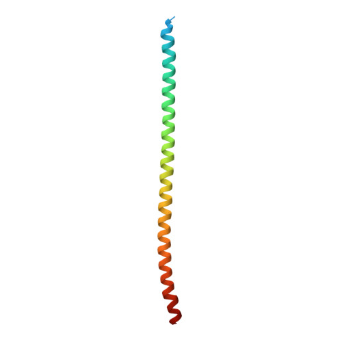
8FXF

Crystal structure of the coiled-coil domain of TRIM56


Experimental Data Snapshot

  • Method: X-RAY DIFFRACTION
  • Resolution: 2.80 Å
  • R-Value Free: 0.264 
  • R-Value Work: 0.219 

wwPDB Validation   3D Report Full Report


This is version 1.0 of the entry. See complete history


Literature

TRIM56 coiled-coil domain structure provides insights into its E3 ligase functions.

Lou, X.Ma, B.Zhuang, Y.Xiao, X.Minze, L.J.Xing, J.Zhang, Z.Li, X.C.

(2023) Comput Struct Biotechnol J 21: 2801-2808

  • DOI: https://doi.org/10.1016/j.csbj.2023.04.022
  • Primary Citation of Related Structures:  
    8FXF

  • PubMed Abstract: 

    Protein ubiquitination is a post-translation modification mediated by E3 ubiquitin ligases. The RING domain E3 ligases are the largest family of E3 ubiquitin ligases, they act as a scaffold, bringing the E2-ubiquitin complex and its substrate together to facilitate direct ubiquitin transfer. However, the quaternary structures of RING E3 ligases that perform ubiquitin transfer remain poorly understood. In this study, we solved the crystal structure of TRIM56, a member of the RING E3 ligase. The structure of the coiled-coil domain indicated that the two anti-parallel dimers bound together to form a tetramer at a small crossing angle. This tetramer structure allows two RING domains to exist on each side to form an active homodimer in supporting ubiquitin transfer from E2 to its nearby substrate recruited by the C-terminal domains on the same side. These findings suggest that the coiled-coil domain-mediated tetramer is a feasible scaffold for facilitating the recruitment and transfer of ubiquitin to accomplish E3 ligase activity.


  • Organizational Affiliation

    Immunobiology and Transplant Science Center and Department of Surgery, Houston Methodist Research Institute, Houston, TX, USA.


Macromolecules
Find similar proteins by:  (by identity cutoff)  |  3D Structure
Entity ID: 1
MoleculeChains Sequence LengthOrganismDetailsImage
E3 ubiquitin-protein ligase TRIM56
A, B, C, D
93Mus musculusMutation(s): 0 
Gene Names: Trim56
EC: 2.3.2.27
UniProt & NIH Common Fund Data Resources
Find proteins for Q80VI1 (Mus musculus)
Explore Q80VI1 
Go to UniProtKB:  Q80VI1
IMPC:  MGI:2685298
Entity Groups  
Sequence Clusters30% Identity50% Identity70% Identity90% Identity95% Identity100% Identity
UniProt GroupQ80VI1
Sequence Annotations
Expand
  • Reference Sequence
Small Molecules
Ligands 1 Unique
IDChains Name / Formula / InChI Key2D Diagram3D Interactions
EPE
Query on EPE

Download Ideal Coordinates CCD File 
E [auth D]4-(2-HYDROXYETHYL)-1-PIPERAZINE ETHANESULFONIC ACID
C8 H18 N2 O4 S
JKMHFZQWWAIEOD-UHFFFAOYSA-N
Experimental Data & Validation

Experimental Data

  • Method: X-RAY DIFFRACTION
  • Resolution: 2.80 Å
  • R-Value Free: 0.264 
  • R-Value Work: 0.219 
  • Space Group: I 41
Unit Cell:
Length ( Å )Angle ( ˚ )
a = 136.679α = 90
b = 136.679β = 90
c = 65.27γ = 90
Software Package:
Software NamePurpose
REFMACrefinement
MOSFLMdata reduction
Aimlessdata scaling
PHENIXphasing

Structure Validation

View Full Validation Report


Entry History & Funding Information

Deposition Data


Funding OrganizationLocationGrant Number
National Institutes of Health/National Institute Of Allergy and Infectious Diseases (NIH/NIAID)United StatesR01 AI080779
National Institutes of Health/National Institute Of Allergy and Infectious Diseases (NIH/NIAID)United StatesR01AI155488

Revision History  (Full details and data files)

  • Version 1.0: 2023-05-24
    Type: Initial release