8F8Y

PHF2 (PHD+JMJ) in Complex with VRK1 N-Terminal Peptide


Experimental Data Snapshot

  • Method: X-RAY DIFFRACTION
  • Resolution: 3.06 Å
  • R-Value Free: 0.249 
  • R-Value Work: 0.195 
  • R-Value Observed: 0.197 

wwPDB Validation   3D Report Full Report


This is version 1.1 of the entry. See complete history


Literature

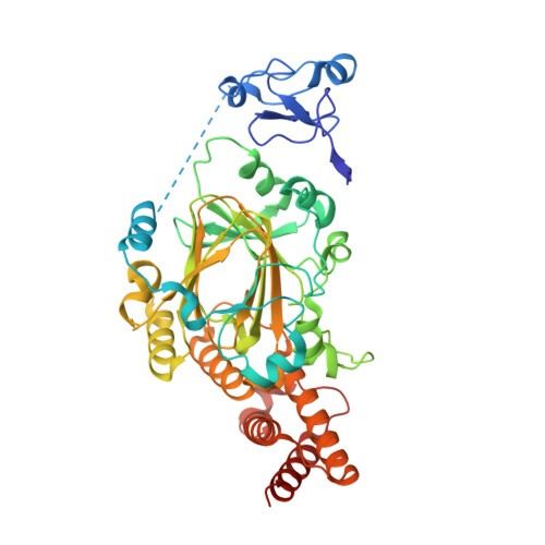
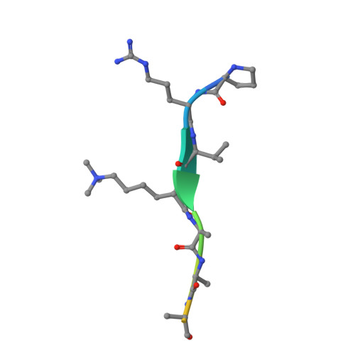
A complete methyl-lysine binding aromatic cage constructed by two domains of PHF2.

Horton, J.R.Zhou, J.Chen, Q.Zhang, X.Bedford, M.T.Cheng, X.

(2022) J Biol Chem 299: 102862-102862

  • DOI: https://doi.org/10.1016/j.jbc.2022.102862
  • Primary Citation of Related Structures:  
    8F8Y, 8F8Z

  • PubMed Abstract: 

    The N-terminal half of PHF2 harbors both a plant homeodomain (PHD) and a Jumonji domain. The PHD recognizes both histone H3 trimethylated at lysine 4 and methylated nonhistone proteins including vaccinia-related kinase 1 (VRK1). The Jumonji domain erases the repressive dimethylation mark from histone H3 lysine 9 (H3K9me2) at select promoters. The N-terminal amino acid sequences of H3 (AR 2 TK 4 ) and VRK1 (PR 2 VK 4 ) bear an arginine at position 2 and lysine at position 4. Here, we show that the PHF2 N-terminal half binds to H3 and VRK1 peptides containing K4me3, with dissociation constants (K D values) of 160 nM and 42 nM, respectively, which are 4 × and 21 × lower (and higher affinities) than for the isolated PHD domain of PHF2. X-ray crystallography revealed that the K4me3-containing peptide is positioned within the PHD and Jumonji interface, with the positively charged R2 residue engaging acidic residues of the PHD and Jumonji domains and with the K4me3 moiety encircled by aromatic residues from both domains. We suggest that the micromolar binding affinities commonly observed for isolated methyl-lysine reader domains could be improved via additional functional interactions within the same polypeptide or its binding partners.


  • Organizational Affiliation

    Department of Epigenetics and Molecular Carcinogenesis, University of Texas MD Anderson Cancer Center, Houston, Texas, USA.


Macromolecules
Find similar proteins by:  (by identity cutoff)  |  3D Structure
Entity ID: 1
MoleculeChains Sequence LengthOrganismDetailsImage
Lysine-specific demethylase PHF2
A, B
455Homo sapiensMutation(s): 0 
Gene Names: PHF2CENP-35KIAA0662
EC: 1.14.11
UniProt & NIH Common Fund Data Resources
Find proteins for O75151 (Homo sapiens)
Explore O75151 
Go to UniProtKB:  O75151
PHAROS:  O75151
GTEx:  ENSG00000197724 
Entity Groups  
Sequence Clusters30% Identity50% Identity70% Identity90% Identity95% Identity100% Identity
UniProt GroupO75151
Sequence Annotations
Expand
  • Reference Sequence

Find similar proteins by:  Sequence   |   3D Structure  

Entity ID: 2
MoleculeChains Sequence LengthOrganismDetailsImage
Serine/threonine-protein kinase VRK1 N-terminus peptideC [auth E],
D [auth F]
12Homo sapiensMutation(s): 0 
EC: 2.7.11.1
UniProt & NIH Common Fund Data Resources
Find proteins for Q99986 (Homo sapiens)
Explore Q99986 
Go to UniProtKB:  Q99986
PHAROS:  Q99986
GTEx:  ENSG00000100749 
Entity Groups  
Sequence Clusters30% Identity50% Identity70% Identity90% Identity95% Identity100% Identity
UniProt GroupQ99986
Sequence Annotations
Expand
  • Reference Sequence
Small Molecules
Ligands 3 Unique
IDChains Name / Formula / InChI Key2D Diagram3D Interactions
SO4
Query on SO4

Download Ideal Coordinates CCD File 
AA [auth B]
BA [auth B]
CA [auth B]
DA [auth B]
G [auth A]
AA [auth B],
BA [auth B],
CA [auth B],
DA [auth B],
G [auth A],
H [auth A],
I [auth A],
J [auth A],
K [auth A],
L [auth A],
M [auth A],
N [auth A],
O [auth A],
P [auth A],
T [auth B],
U [auth B],
V [auth B],
W [auth B],
X [auth B],
Y [auth B]
SULFATE ION
O4 S
QAOWNCQODCNURD-UHFFFAOYSA-L
ZN
Query on ZN

Download Ideal Coordinates CCD File 
E [auth A],
F [auth A],
Q [auth B],
R [auth B]
ZINC ION
Zn
PTFCDOFLOPIGGS-UHFFFAOYSA-N
EDO
Query on EDO

Download Ideal Coordinates CCD File 
S [auth B],
Z [auth B]
1,2-ETHANEDIOL
C2 H6 O2
LYCAIKOWRPUZTN-UHFFFAOYSA-N
Modified Residues  1 Unique
IDChains TypeFormula2D DiagramParent
M3L
Query on M3L
C [auth E],
D [auth F]
L-PEPTIDE LINKINGC9 H21 N2 O2LYS
Experimental Data & Validation

Experimental Data

  • Method: X-RAY DIFFRACTION
  • Resolution: 3.06 Å
  • R-Value Free: 0.249 
  • R-Value Work: 0.195 
  • R-Value Observed: 0.197 
  • Space Group: P 1 21 1
Unit Cell:
Length ( Å )Angle ( ˚ )
a = 74.406α = 90
b = 84.065β = 103.99
c = 105.215γ = 90
Software Package:
Software NamePurpose
SERGUIdata collection
PHENIXrefinement
HKL-2000data reduction
HKL-2000data scaling
PHENIXphasing

Structure Validation

View Full Validation Report


Entry History & Funding Information

Deposition Data


Funding OrganizationLocationGrant Number
National Institutes of Health/National Institute of General Medical Sciences (NIH/NIGMS)United StatesR35GM134744
Cancer Prevention and Research Institute of Texas (CPRIT)United StatesRR160029

Revision History  (Full details and data files)

  • Version 1.0: 2023-01-18
    Type: Initial release
  • Version 1.1: 2023-02-08
    Changes: Database references