Variability in phenylalanine side chain conformations facilitates broad substrate tolerance of fatty acid binding in cockroach milk proteins.
Santhakumari, P.R., Dhanabalan, K., Virani, S., Hopf-Jannasch, A.S., Benoit, J.B., Chopra, G., Subramanian, R.(2023) PLoS One 18: e0280009-e0280009
- PubMed: 37384723 Search on PubMedSearch on PubMed Central
- DOI: https://doi.org/10.1371/journal.pone.0280009
- Primary Citation Related Structures:
8F0V, 8F0Y - PubMed Abstract:
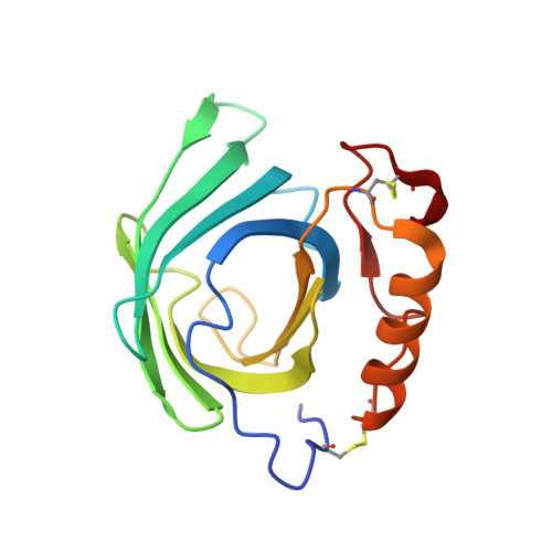
Diploptera punctata, also known as the Pacific beetle cockroach, is a viviparous cockroach that gives birth to live offspring and secretes a highly concentrated mixture of glycosylated proteins as a source of nourishment for developing embryos. These proteins are lipocalins that bind to lipids and crystallize in the gut of the embryo. A structure of milk crystals harvested from the embryos showed that the milk-derived crystals were heterogeneous and made of three proteins (called Lili-Mips). We hypothesized that the isoforms of Lili-Mip would display different affinities for fatty acids due to the ability of the pocket to bind multiple acyl chain lengths. We previously reported the structures of Lili-Mip from crystals grown in vivo and recombinantly expressed Lili-Mip2. These structures are similar, and both bind to several fatty acids. This study explores the specificity and affinity of fatty acid binding to recombinantly expressed Lili-Mip 1, 2 & 3. We show that all isoforms can bind to different fatty acids with similar affinities. We also report the thermostability of Lili-Mip is pH dependent, where stability is highest at acidic pH and declines as the pH increases to physiological levels near 7.0. We show that thermostability is an inherent property of the protein, and glycosylation and ligand binding do not change it significantly. Measuring the pH in the embryo's gut lumen and gut cells suggests that the pH in the gut is acidic and the pH inside the gut cells is closer to neutral pH. In various crystal structures (reported here and previously by us), Phe-98 and Phe-100 occupy multiple conformations in the binding pocket. In our earlier work, we had shown that the loops at the entrance could adapt various conformations to change the size of the binding pocket. Here we show Phe-98 and Phe-100 can reorient to stabilize interactions at the bottom of the cavity-and change the volume of the cavity from 510 Å3 to 337 Å3. Together they facilitate the binding of fatty acids of different acyl chain lengths.
- Institute for Stem Cell Science and Regenerative Medicine, Bengaluru, Karnataka, India.
Organizational Affiliation: