8ESV

Structure of human ADAM10-Tspan15 complex bound to 11G2 vFab


Experimental Data Snapshot

  • Method: ELECTRON MICROSCOPY
  • Resolution: 3.30 Å
  • Aggregation State: PARTICLE 
  • Reconstruction Method: SINGLE PARTICLE 

wwPDB Validation   3D Report Full Report


This is version 1.3 of the entry. See complete history


Literature

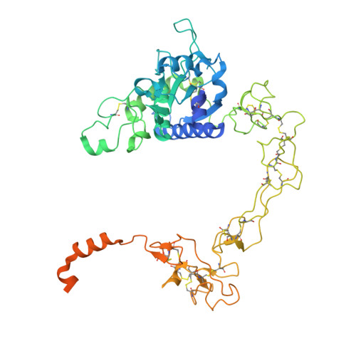
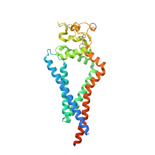
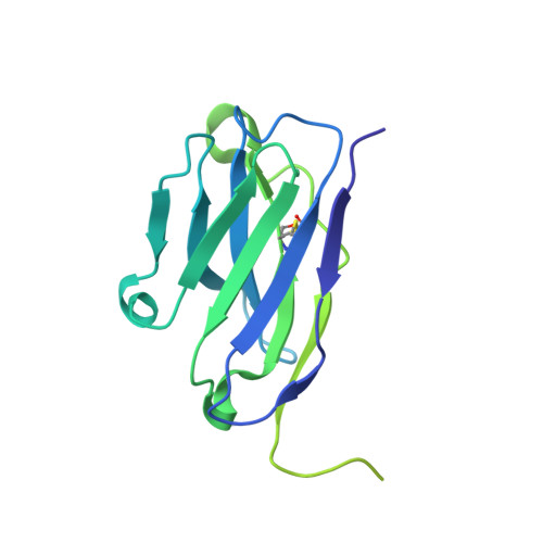
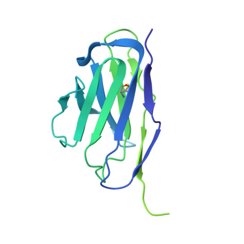
Structural basis for membrane-proximal proteolysis of substrates by ADAM10.

Lipper, C.H.Egan, E.D.Gabriel, K.H.Blacklow, S.C.

(2023) Cell 186: 3632-3641.e10

  • DOI: https://doi.org/10.1016/j.cell.2023.06.026
  • Primary Citation of Related Structures:  
    8ESV

  • PubMed Abstract: 

    The endopeptidase ADAM10 is a critical catalyst for the regulated proteolysis of key drivers of mammalian development, physiology, and non-amyloidogenic cleavage of APP as the primary α-secretase. ADAM10 function requires the formation of a complex with a C8-tetraspanin protein, but how tetraspanin binding enables positioning of the enzyme active site for membrane-proximal cleavage remains unknown. We present here a cryo-EM structure of a vFab-ADAM10-Tspan15 complex, which shows that Tspan15 binding relieves ADAM10 autoinhibition and acts as a molecular measuring stick to position the enzyme active site about 20 Å from the plasma membrane for membrane-proximal substrate cleavage. Cell-based assays of N-cadherin shedding establish that the positioning of the active site by the interface between the ADAM10 catalytic domain and the bound tetraspanin influences selection of the preferred cleavage site. Together, these studies reveal the molecular mechanism underlying ADAM10 proteolysis at membrane-proximal sites and offer a roadmap for its modulation in disease.


  • Organizational Affiliation

    Department of Biological Chemistry and Molecular Pharmacology, Blavatnik Institute, Harvard Medical School, Boston, MA 02115, USA.


Macromolecules
Find similar proteins by:  (by identity cutoff)  |  3D Structure
Entity ID: 1
MoleculeChains Sequence LengthOrganismDetailsImage
Disintegrin and metalloproteinase domain-containing protein 10543Homo sapiensMutation(s): 0 
Gene Names: ADAM10KUZMADM
EC: 3.4.24.81
Membrane Entity: Yes 
UniProt & NIH Common Fund Data Resources
Find proteins for O14672 (Homo sapiens)
Explore O14672 
Go to UniProtKB:  O14672
PHAROS:  O14672
GTEx:  ENSG00000137845 
Entity Groups  
Sequence Clusters30% Identity50% Identity70% Identity90% Identity95% Identity100% Identity
UniProt GroupO14672
Sequence Annotations
Expand
  • Reference Sequence
Find similar proteins by:  (by identity cutoff)  |  3D Structure
Entity ID: 2
MoleculeChains Sequence LengthOrganismDetailsImage
Tetraspanin-15306Homo sapiensMutation(s): 0 
Gene Names: TSPAN15NET7TM4SF15UNQ677/PRO1311
Membrane Entity: Yes 
UniProt & NIH Common Fund Data Resources
Find proteins for O95858 (Homo sapiens)
Explore O95858 
Go to UniProtKB:  O95858
PHAROS:  O95858
GTEx:  ENSG00000099282 
Entity Groups  
Sequence Clusters30% Identity50% Identity70% Identity90% Identity95% Identity100% Identity
UniProt GroupO95858
Sequence Annotations
Expand
  • Reference Sequence
Find similar proteins by:  (by identity cutoff)  |  3D Structure
Entity ID: 3
MoleculeChains Sequence LengthOrganismDetailsImage
11G2 Fab Heavy ChainC [auth H]225Mus musculusMutation(s): 0 
Entity Groups  
Sequence Clusters30% Identity50% Identity70% Identity90% Identity95% Identity100% Identity
Sequence Annotations
Expand
  • Reference Sequence
Find similar proteins by:  (by identity cutoff)  |  3D Structure
Entity ID: 4
MoleculeChains Sequence LengthOrganismDetailsImage
11G2 Fab Light ChainD [auth L]221Mus musculusMutation(s): 0 
Entity Groups  
Sequence Clusters30% Identity50% Identity70% Identity90% Identity95% Identity100% Identity
Sequence Annotations
Expand
  • Reference Sequence
Oligosaccharides

Help

Entity ID: 5
MoleculeChains Length2D Diagram Glycosylation3D Interactions
alpha-D-mannopyranose-(1-3)-alpha-D-mannopyranose-(1-4)-2-acetamido-2-deoxy-beta-D-glucopyranose-(1-4)-2-acetamido-2-deoxy-beta-D-glucopyranoseE [auth C]4N-Glycosylation
Glycosylation Resources
GlyTouCan:  G48068RF
GlyCosmos:  G48068RF
GlyGen:  G48068RF
Entity ID: 6
MoleculeChains Length2D Diagram Glycosylation3D Interactions
alpha-D-mannopyranose-(1-4)-2-acetamido-2-deoxy-beta-D-glucopyranose-(1-4)-2-acetamido-2-deoxy-beta-D-glucopyranoseF [auth D]3N-Glycosylation
Glycosylation Resources
GlyTouCan:  G62182OO
GlyCosmos:  G62182OO
GlyGen:  G62182OO
Small Molecules
Ligands 5 Unique
IDChains Name / Formula / InChI Key2D Diagram3D Interactions
Y01
Query on Y01

Download Ideal Coordinates CCD File 
K [auth B],
L [auth B]
CHOLESTEROL HEMISUCCINATE
C31 H50 O4
WLNARFZDISHUGS-MIXBDBMTSA-N
BAT
Query on BAT

Download Ideal Coordinates CCD File 
J [auth A]4-(N-HYDROXYAMINO)-2R-ISOBUTYL-2S-(2-THIENYLTHIOMETHYL)SUCCINYL-L-PHENYLALANINE-N-METHYLAMIDE
C23 H31 N3 O4 S2
XFILPEOLDIKJHX-QYZOEREBSA-N
NAG
Query on NAG

Download Ideal Coordinates CCD File 
G [auth A]2-acetamido-2-deoxy-beta-D-glucopyranose
C8 H15 N O6
OVRNDRQMDRJTHS-FMDGEEDCSA-N
ZN
Query on ZN

Download Ideal Coordinates CCD File 
I [auth A]ZINC ION
Zn
PTFCDOFLOPIGGS-UHFFFAOYSA-N
CA
Query on CA

Download Ideal Coordinates CCD File 
H [auth A]CALCIUM ION
Ca
BHPQYMZQTOCNFJ-UHFFFAOYSA-N
Experimental Data & Validation

Experimental Data

  • Method: ELECTRON MICROSCOPY
  • Resolution: 3.30 Å
  • Aggregation State: PARTICLE 
  • Reconstruction Method: SINGLE PARTICLE 
EM Software:
TaskSoftware PackageVersion
MODEL REFINEMENTPHENIX
RECONSTRUCTIONcryoSPARC3.3.1

Structure Validation

View Full Validation Report


Entry History & Funding Information

Deposition Data


Funding OrganizationLocationGrant Number
National Institutes of Health/National Cancer Institute (NIH/NCI)United StatesR35 CA220340
Other privateGift from Edward B. Goodnow

Revision History  (Full details and data files)

  • Version 1.0: 2023-06-14
    Type: Initial release
  • Version 1.1: 2023-08-09
    Changes: Database references
  • Version 1.2: 2023-08-16
    Changes: Data collection
  • Version 1.3: 2023-08-30
    Changes: Database references