8DKT

Crystal Structure of Septin1 - Septin2 heterocomplex from Drosophila melanogaster


Experimental Data Snapshot

  • Method: X-RAY DIFFRACTION
  • Resolution: 2.38 Å
  • R-Value Free: 0.255 
  • R-Value Work: 0.217 
  • R-Value Observed: 0.219 

wwPDB Validation   3D Report Full Report


Ligand Structure Quality Assessment 


This is version 1.2 of the entry. See complete history


Literature

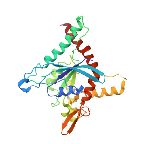
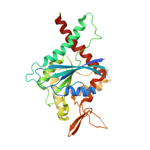
Conservation and divergence of the G-interfaces of Drosophila melanogaster septins.

de Freitas Fernandes, A.Leonardo, D.A.Cavini, I.A.Rosa, H.V.D.Vargas, J.A.D'Muniz Pereira, H.Nascimento, A.S.Garratt, R.C.

(2023) Cytoskeleton (Hoboken) 80: 153-168

  • DOI: https://doi.org/10.1002/cm.21740
  • Primary Citation of Related Structures:  
    8DKT

  • PubMed Abstract: 

    Septins possess a conserved guanine nucleotide-binding (G) domain that participates in the stabilization of organized hetero-oligomeric complexes which assemble into filaments, rings and network-like structures. The fruit fly, Drosophila melanogaster, has five such septin genes encoding Sep1, Sep2, Sep4, Sep5 and Pnut. Here, we report the crystal structure of the heterodimer formed between the G-domains of Sep1 and Sep2, the first from an insect to be described to date. A G-interface stabilizes the dimer (in agreement with the expected arrangement for the Drosophila hexameric particle) and this bears significant resemblance to its human counterparts, even down to the level of individual amino acid interactions. On the other hand, a model for the G-interface formed between the two copies of Pnut which occupy the centre of the hexamer, shows important structural differences, including the loss of a highly favourable bifurcated salt-bridge network. Whereas wild-type Pnut purifies as a monomer, the reintroduction of the salt-bridge network results in stabilizing the dimeric interface in solution as shown by size exclusion chromatography and thermal stability measurements. Adaptive steered molecular dynamics reveals an unzipping mechanism for dimer dissociation which initiates at a point of electrostatic repulsion within the switch II region. Overall, the data contribute to a better understanding of the molecular interactions involved in septin assembly/disassembly.


  • Organizational Affiliation

    São Carlos Institute of Physics, University of São Paulo, São Carlos, Brazil.


Macromolecules
Find similar proteins by:  (by identity cutoff)  |  3D Structure
Entity ID: 1
MoleculeChains Sequence LengthOrganismDetailsImage
Septin-1275Drosophila melanogasterMutation(s): 0 
Gene Names: Sep1Diff6ibyCG1403
UniProt
Find proteins for P42207 (Drosophila melanogaster)
Explore P42207 
Go to UniProtKB:  P42207
Entity Groups  
Sequence Clusters30% Identity50% Identity70% Identity90% Identity95% Identity100% Identity
UniProt GroupP42207
Sequence Annotations
Expand
  • Reference Sequence
Find similar proteins by:  (by identity cutoff)  |  3D Structure
Entity ID: 2
MoleculeChains Sequence LengthOrganismDetailsImage
Septin-2282Drosophila melanogasterMutation(s): 0 
Gene Names: Sep2CG4173
UniProt
Find proteins for P54359 (Drosophila melanogaster)
Explore P54359 
Go to UniProtKB:  P54359
Entity Groups  
Sequence Clusters30% Identity50% Identity70% Identity90% Identity95% Identity100% Identity
UniProt GroupP54359
Sequence Annotations
Expand
  • Reference Sequence
Experimental Data & Validation

Experimental Data

  • Method: X-RAY DIFFRACTION
  • Resolution: 2.38 Å
  • R-Value Free: 0.255 
  • R-Value Work: 0.217 
  • R-Value Observed: 0.219 
  • Space Group: C 2 2 21
Unit Cell:
Length ( Å )Angle ( ˚ )
a = 73.577α = 90
b = 149.423β = 90
c = 155.34γ = 90
Software Package:
Software NamePurpose
PHASERphasing
PHENIXrefinement
PDB_EXTRACTdata extraction
autoPROCdata reduction
autoPROCdata scaling

Structure Validation

View Full Validation Report


Ligand Structure Quality Assessment 


Entry History & Funding Information

Deposition Data


Funding OrganizationLocationGrant Number
Sao Paulo Research Foundation (FAPESP)Brazil2020/02897-1

Revision History  (Full details and data files)

  • Version 1.0: 2023-01-11
    Type: Initial release
  • Version 1.1: 2023-10-04
    Changes: Data collection, Database references
  • Version 1.2: 2023-10-25
    Changes: Refinement description