Structure-function studies reveal ComEA contains an oligomerization domain essential for transformation in gram-positive bacteria.
Ahmed, I., Hahn, J., Henrickson, A., Khaja, F.T., Demeler, B., Dubnau, D., Neiditch, M.B.(2022) Nat Commun 13: 7724-7724
- PubMed: 36513643 Search on PubMedSearch on PubMed Central
- DOI: https://doi.org/10.1038/s41467-022-35129-0
- Primary Citation Related Structures:
8DFK, 8DSS - PubMed Abstract:
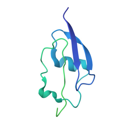
An essential step in bacterial transformation is the uptake of DNA into the periplasm, across the thick peptidoglycan cell wall of Gram-positive bacteria, or the outer membrane and thin peptidoglycan layer of Gram-negative bacteria. ComEA, a DNA-binding protein widely conserved in transformable bacteria, is required for this uptake step. Here we determine X-ray crystal structures of ComEA from two Gram-positive species, Bacillus subtilis and Geobacillus stearothermophilus, identifying a domain that is absent in Gram-negative bacteria. X-ray crystallographic, genetic, and analytical ultracentrifugation (AUC) analyses reveal that this domain drives ComEA oligomerization, which we show is required for transformation. We use multi-wavelength AUC (MW-AUC) to characterize the interaction between DNA and the ComEA DNA-binding domain. Finally, we present a model for the interaction of the ComEA DNA-binding domain with DNA, suggesting that ComEA oligomerization may provide a pulling force that drives DNA uptake across the thick cell walls of Gram-positive bacteria.
- Department of Microbiology, Biochemistry, and Molecular Genetics, New Jersey Medical School, Rutgers Biomedical Health Sciences, Newark, NJ, 07103, USA.
Organizational Affiliation: