8DC7

Crystal structure of p53 Y220C covalently bound to indole KG10


Experimental Data Snapshot

  • Method: X-RAY DIFFRACTION
  • Resolution: 1.99 Å
  • R-Value Free: 0.193 
  • R-Value Work: 0.160 
  • R-Value Observed: 0.162 

wwPDB Validation   3D Report Full Report


Ligand Structure Quality Assessment 


This is version 1.2 of the entry. See complete history


Literature

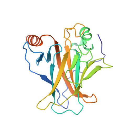
A Small Molecule Reacts with the p53 Somatic Mutant Y220C to Rescue Wild-type Thermal Stability.

Guiley, K.Z.Shokat, K.M.

(2023) Cancer Discov 13: 56-69

  • DOI: https://doi.org/10.1158/2159-8290.CD-22-0381
  • Primary Citation of Related Structures:  
    8DC4, 8DC6, 8DC7, 8DC8

  • PubMed Abstract: 

    The transcription factor and tumor suppressor protein p53 is the most frequently mutated and inactivated gene in cancer. Mutations in p53 result in deregulated cell proliferation and genomic instability, both hallmarks of cancer. There are currently no therapies available that directly target mutant p53 to rescue wild-type function. In this study, we identify covalent compsounds that selectively react with the p53 somatic mutant cysteine Y220C and restore wild-type thermal stability.


  • Organizational Affiliation

    Department of Cellular and Molecular Pharmacology and Howard Hughes Medical Institute, University of California, San Francisco, San Francisco, California.


Macromolecules
Find similar proteins by:  (by identity cutoff)  |  3D Structure
Entity ID: 1
MoleculeChains Sequence LengthOrganismDetailsImage
Cellular tumor antigen p53222Homo sapiensMutation(s): 1 
Gene Names: TP53P53
UniProt & NIH Common Fund Data Resources
Find proteins for P04637 (Homo sapiens)
Explore P04637 
Go to UniProtKB:  P04637
PHAROS:  P04637
GTEx:  ENSG00000141510 
Entity Groups  
Sequence Clusters30% Identity50% Identity70% Identity90% Identity95% Identity100% Identity
UniProt GroupP04637
Sequence Annotations
Expand
  • Reference Sequence
Small Molecules
Ligands 4 Unique
IDChains Name / Formula / InChI Key2D Diagram3D Interactions
R4R (Subject of Investigation/LOI)
Query on R4R

Download Ideal Coordinates CCD File 
H [auth A]4-[4-(4-methylpiperazin-1-yl)phenyl]-1-(2-methylprop-2-enoyl)-1H-indole-3-carbaldehyde, bound form
C24 H27 N3 O2
UKTLOAMGFGATOJ-UHFFFAOYSA-N
SO4
Query on SO4

Download Ideal Coordinates CCD File 
B [auth A],
C [auth A],
D [auth A]
SULFATE ION
O4 S
QAOWNCQODCNURD-UHFFFAOYSA-L
ZN (Subject of Investigation/LOI)
Query on ZN

Download Ideal Coordinates CCD File 
E [auth A]ZINC ION
Zn
PTFCDOFLOPIGGS-UHFFFAOYSA-N
MG
Query on MG

Download Ideal Coordinates CCD File 
F [auth A],
G [auth A]
MAGNESIUM ION
Mg
JLVVSXFLKOJNIY-UHFFFAOYSA-N
Experimental Data & Validation

Experimental Data

  • Method: X-RAY DIFFRACTION
  • Resolution: 1.99 Å
  • R-Value Free: 0.193 
  • R-Value Work: 0.160 
  • R-Value Observed: 0.162 
  • Space Group: C 2 2 21
Unit Cell:
Length ( Å )Angle ( ˚ )
a = 68.66α = 90
b = 85.92β = 90
c = 84.04γ = 90
Software Package:
Software NamePurpose
Blu-Icedata collection
iMOSFLMdata reduction
Aimlessdata scaling
PHASERphasing
PHENIXrefinement

Structure Validation

View Full Validation Report


Ligand Structure Quality Assessment 


Entry History & Funding Information

Deposition Data


Funding OrganizationLocationGrant Number
Howard Hughes Medical Institute (HHMI)United StatesDRG: 2399-20

Revision History  (Full details and data files)

  • Version 1.0: 2022-10-12
    Type: Initial release
  • Version 1.1: 2023-01-18
    Changes: Database references
  • Version 1.2: 2023-10-25
    Changes: Data collection, Refinement description