High pressure freezing and cryo-sectioning can be used for protein structure determination by electron diffraction.
Moriscot, C., Schoehn, G., Housset, D.(2023) Ultramicroscopy 254: 113834-113834
- PubMed: 37666105 Search on PubMed
- DOI: https://doi.org/10.1016/j.ultramic.2023.113834
- Primary Citation Related Structures:
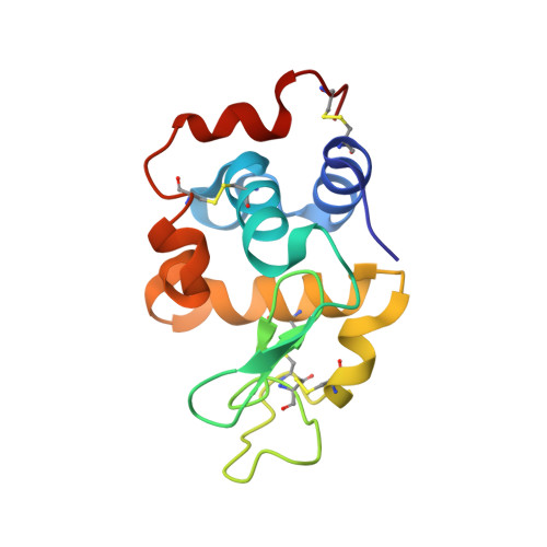
8CPC - PubMed Abstract:
Electron diffraction of three-dimensional nanometer sized crystals has emerged since 2013 as an efficient technique to solve the structure of both small organic molecules and model proteins. However, the major bottleneck of the technique when applied to protein samples is to produce nano-crystals that do not exceed 200 to 300 nm in at least one dimension and to deposit them on a grid while keeping the minimum amount of solvent around them. Since the presence of amorphous solvent around the crystal, necessary to preserve its integrity, increases the amount of diffuse scattering, thus degrading the signal-to noise ratio of the diffraction signal, other sample preparation strategies have been developed. One of them is the milling of thin crystal lamella using focused ion beam (FIB), which was successfully applied to several protein crystals. Here, we present a new approach that uses cryo-sectioning after high pressure freezing of dextran embedded protein crystals. 150 to 200 nm thick cryo-sections of hen egg white lysozyme tetragonal crystals where used for electron diffraction experiments. Complete diffraction data up to 2.9 Å resolution have been collected and the lysozyme structure has been solved by molecular replacement and refined against these data. Our data demonstrate that cryo-sectioning can preserve protein structure at high resolution and can be used as a new sample preparation technique for 3D electron diffraction experiments of protein crystals. The different orientations found in the crystal chips and their large number resulting from the cryo-sectioning make the latter an attractive approach as it combines advantages from both blotting approaches (number of crystals) and FIB-milling (controlled thickness and absence of solvent around the crystal).
- Univ. Grenoble Alpes, CNRS, CEA, ISBG, IBS, F-38000 Grenoble, France. Electronic address: christine.moriscot@ibs.fr.
Organizational Affiliation: