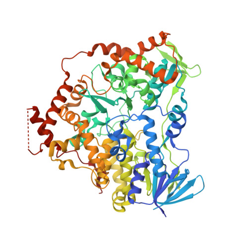
8CJD

AetF, a single-component flavin-dependent tryptophan halogenase


Experimental Data Snapshot

  • Method: X-RAY DIFFRACTION
  • Resolution: 1.70 Å
  • R-Value Free: 0.185 
  • R-Value Work: 0.159 
  • R-Value Observed: 0.160 

Starting Model: in silico
View more details

wwPDB Validation   3D Report Full Report


Ligand Structure Quality Assessment 


This is version 1.3 of the entry. See complete history


Literature

Structural basis of regioselective tryptophan dibromination by the single-component flavin-dependent halogenase AetF.

Gafe, S.Niemann, H.H.

(2023) Acta Crystallogr D Struct Biol 79: 596-609

  • DOI: https://doi.org/10.1107/S2059798323004254
  • Primary Citation of Related Structures:  
    8CJD, 8CJE, 8CJF, 8CJG

  • PubMed Abstract: 

    The flavin-dependent halogenase (FDH) AetF successively brominates tryptophan at C5 and C7 to generate 5,7-dibromotryptophan. In contrast to the well studied two-component tryptophan halogenases, AetF is a single-component flavoprotein monooxygenase. Here, crystal structures of AetF alone and in complex with various substrates are presented, representing the first experimental structures of a single-component FDH. Rotational pseudosymmetry and pseudomerohedral twinning complicated the phasing of one structure. AetF is structurally related to flavin-dependent monooxygenases. It contains two dinucleotide-binding domains for binding the ADP moiety with unusual sequences that deviate from the consensus sequences GXGXXG and GXGXXA. A large domain tightly binds the cofactor flavin adenine dinucleotide (FAD), while the small domain responsible for binding the nicotinamide adenine dinucleotide (NADP) is unoccupied. About half of the protein forms additional structural elements containing the tryptophan binding site. FAD and tryptophan are about 16 Å apart. A tunnel between them presumably allows diffusion of the active halogenating agent hypohalous acid from FAD to the substrate. Tryptophan and 5-bromotryptophan bind to the same site but with a different binding pose. A flip of the indole moiety identically positions C5 of tryptophan and C7 of 5-bromotryptophan next to the tunnel and to catalytic residues, providing a simple explanation for the regioselectivity of the two successive halogenations. AetF can also bind 7-bromotryptophan in the same orientation as tryptophan. This opens the way for the biocatalytic production of differentially dihalogenated tryptophan derivatives. The structural conservation of a catalytic lysine suggests a way to identify novel single-component FDHs.


  • Organizational Affiliation

    Department of Chemistry, Bielefeld University, Universitaetsstrasse 25, 33615 Bielefeld, Germany.


Macromolecules
Find similar proteins by:  (by identity cutoff)  |  3D Structure
Entity ID: 1
MoleculeChains Sequence LengthOrganismDetailsImage
AetF
A, B
663Aetokthonos hydrillicola Thurmond2011Mutation(s): 0 
Gene Names: aetF
UniProt
Find proteins for A0A861B9Z9 (Aetokthonos hydrillicola Thurmond2011)
Explore A0A861B9Z9 
Go to UniProtKB:  A0A861B9Z9
Entity Groups  
Sequence Clusters30% Identity50% Identity70% Identity90% Identity95% Identity100% Identity
UniProt GroupA0A861B9Z9
Sequence Annotations
Expand
  • Reference Sequence
Small Molecules
Ligands 5 Unique
IDChains Name / Formula / InChI Key2D Diagram3D Interactions
FAD (Subject of Investigation/LOI)
Query on FAD

Download Ideal Coordinates CCD File 
C [auth A],
G [auth B]
FLAVIN-ADENINE DINUCLEOTIDE
C27 H33 N9 O15 P2
VWWQXMAJTJZDQX-UYBVJOGSSA-N
PEG
Query on PEG

Download Ideal Coordinates CCD File 
E [auth A],
I [auth B]
DI(HYDROXYETHYL)ETHER
C4 H10 O3
MTHSVFCYNBDYFN-UHFFFAOYSA-N
EDO
Query on EDO

Download Ideal Coordinates CCD File 
D [auth A],
H [auth B]
1,2-ETHANEDIOL
C2 H6 O2
LYCAIKOWRPUZTN-UHFFFAOYSA-N
CA
Query on CA

Download Ideal Coordinates CCD File 
K [auth B]CALCIUM ION
Ca
BHPQYMZQTOCNFJ-UHFFFAOYSA-N
CL
Query on CL

Download Ideal Coordinates CCD File 
F [auth A],
J [auth B]
CHLORIDE ION
Cl
VEXZGXHMUGYJMC-UHFFFAOYSA-M
Experimental Data & Validation

Experimental Data

Unit Cell:
Length ( Å )Angle ( ˚ )
a = 123.79α = 90
b = 123.79β = 90
c = 88γ = 90
Software Package:
Software NamePurpose
PHENIXrefinement
MxCuBEdata collection
XDSdata reduction
XDSdata scaling
PHASERphasing
Cootmodel building

Structure Validation

View Full Validation Report


Ligand Structure Quality Assessment 


Entry History & Funding Information

Deposition Data


Funding OrganizationLocationGrant Number
Not fundedGermany--

Revision History  (Full details and data files)

  • Version 1.0: 2023-06-14
    Type: Initial release
  • Version 1.1: 2023-06-28
    Changes: Database references
  • Version 1.2: 2023-07-12
    Changes: Database references
  • Version 1.3: 2024-06-19
    Changes: Data collection, Refinement description