8C3C

14-3-3 sigma with Pin1 binding site pS72 and bound Fusicoccin A


Experimental Data Snapshot

  • Method: X-RAY DIFFRACTION
  • Resolution: 1.60 Å
  • R-Value Free: 0.200 
  • R-Value Work: 0.192 
  • R-Value Observed: 0.192 

wwPDB Validation   3D Report Full Report


Ligand Structure Quality Assessment 


This is version 1.1 of the entry. See complete history


Literature

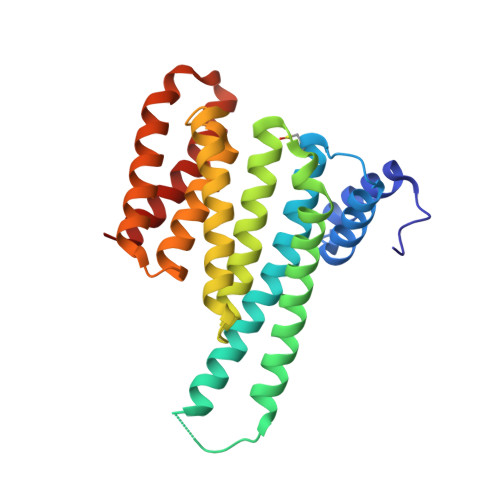
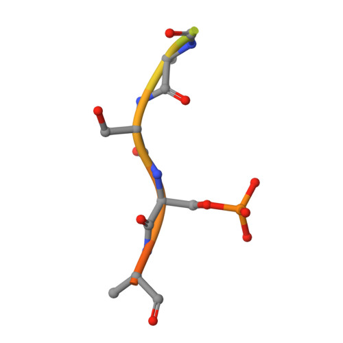
Tracking the mechanism of covalent molecular glue stabilization using native mass spectrometry.

Verhoef, C.J.A.Kay, D.F.van Dijck, L.Doveston, R.G.Brunsveld, L.Leney, A.C.Cossar, P.J.

(2023) Chem Sci 14: 6756-6762

  • DOI: https://doi.org/10.1039/d3sc01732j
  • Primary Citation of Related Structures:  
    8C2G, 8C3C

  • PubMed Abstract: 

    Molecular glues are powerful tools for the control of protein-protein interactions. Yet, the mechanisms underlying multi-component protein complex formation remain poorly understood. Native mass spectrometry (MS) detects multiple protein species simultaneously, providing an entry to elucidate these mechanisms. Here, for the first time, covalent molecular glue stabilization was kinetically investigated by combining native MS with biophysical and structural techniques. This approach elucidated the stoichiometry of a multi-component protein-ligand complex, the assembly order, and the contributions of covalent versus non-covalent binding events that govern molecular glue activity. Aldehyde-based molecular glue activity is initially regulated by cooperative non-covalent binding, followed by slow covalent ligation, further enhancing stabilization. This study provides a framework to investigate the mechanisms of covalent small molecule ligation and informs (covalent) molecular glue development.


  • Organizational Affiliation

    Department of Biomedical Engineering and Institute for Complex Molecular Systems, Eindhoven University of Technology Eindhoven 5600 MB The Netherlands p.cossar@tue.nl.


Macromolecules
Find similar proteins by:  (by identity cutoff)  |  3D Structure
Entity ID: 1
MoleculeChains Sequence LengthOrganismDetailsImage
14-3-3 protein sigma236Homo sapiensMutation(s): 0 
Gene Names: SFNHME1
UniProt & NIH Common Fund Data Resources
Find proteins for P31947 (Homo sapiens)
Explore P31947 
Go to UniProtKB:  P31947
PHAROS:  P31947
GTEx:  ENSG00000175793 
Entity Groups  
Sequence Clusters30% Identity50% Identity70% Identity90% Identity95% Identity100% Identity
UniProt GroupP31947
Sequence Annotations
Expand
  • Reference Sequence

Find similar proteins by:  Sequence   |   3D Structure  

Entity ID: 2
MoleculeChains Sequence LengthOrganismDetailsImage
Peptidyl-prolyl cis-trans isomerase NIMA-interacting 117Homo sapiensMutation(s): 0 
EC: 5.2.1.8
UniProt & NIH Common Fund Data Resources
Find proteins for Q13526 (Homo sapiens)
Explore Q13526 
Go to UniProtKB:  Q13526
PHAROS:  Q13526
GTEx:  ENSG00000127445 
Entity Groups  
Sequence Clusters30% Identity50% Identity70% Identity90% Identity95% Identity100% Identity
UniProt GroupQ13526
Sequence Annotations
Expand
  • Reference Sequence
Small Molecules
Modified Residues  2 Unique
IDChains TypeFormula2D DiagramParent
CSO
Query on CSO
A
L-PEPTIDE LINKINGC3 H7 N O3 SCYS
SEP
Query on SEP
B
L-PEPTIDE LINKINGC3 H8 N O6 PSER
Experimental Data & Validation

Experimental Data

  • Method: X-RAY DIFFRACTION
  • Resolution: 1.60 Å
  • R-Value Free: 0.200 
  • R-Value Work: 0.192 
  • R-Value Observed: 0.192 
  • Space Group: C 2 2 21
Unit Cell:
Length ( Å )Angle ( ˚ )
a = 82.847α = 90
b = 111.544β = 90
c = 62.544γ = 90
Software Package:
Software NamePurpose
Aimlessdata scaling
MOLREPphasing
Cootmodel building
REFMACrefinement
PHENIXrefinement

Structure Validation

View Full Validation Report


Ligand Structure Quality Assessment 


Entry History & Funding Information

Deposition Data


Funding OrganizationLocationGrant Number
Netherlands Organisation for Scientific Research (NWO)Netherlands--

Revision History  (Full details and data files)

  • Version 1.0: 2023-06-14
    Type: Initial release
  • Version 1.1: 2023-07-05
    Changes: Database references