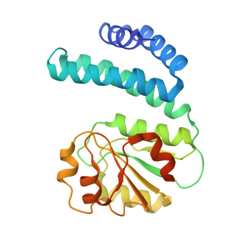
8C37

An intermediate light exposed 2.15 Angstrom crystal structure of H132A variant of cobalamin binding domain belonging to a light-dependent transcription regulator TtCarH obtained under anaerobic conditions


Experimental Data Snapshot

  • Method: X-RAY DIFFRACTION
  • Resolution: 2.15 Å
  • R-Value Free: 0.230 
  • R-Value Work: 0.193 
  • R-Value Observed: 0.195 

wwPDB Validation   3D Report Full Report


Ligand Structure Quality Assessment 


This is version 1.1 of the entry. See complete history


Literature

Redox driven B 12 -ligand switch drives CarH photoresponse.

Poddar, H.Rios-Santacruz, R.Heyes, D.J.Shanmugam, M.Brookfield, A.Johannissen, L.O.Levy, C.W.Jeffreys, L.N.Zhang, S.Sakuma, M.Colletier, J.P.Hay, S.Schiro, G.Weik, M.Scrutton, N.S.Leys, D.

(2023) Nat Commun 14: 5082-5082

  • DOI: https://doi.org/10.1038/s41467-023-40817-6
  • Primary Citation of Related Structures:  
    8C31, 8C32, 8C33, 8C34, 8C35, 8C36, 8C37, 8C73, 8C76

  • PubMed Abstract: 

    CarH is a coenzyme B 12 -dependent photoreceptor involved in regulating carotenoid biosynthesis. How light-triggered cleavage of the B 12 Co-C bond culminates in CarH tetramer dissociation to initiate transcription remains unclear. Here, a series of crystal structures of the CarH B 12 -binding domain after illumination suggest formation of unforeseen intermediate states prior to tetramer dissociation. Unexpectedly, in the absence of oxygen, Co-C bond cleavage is followed by reorientation of the corrin ring and a switch from a lower to upper histidine-Co ligation, corresponding to a pentacoordinate state. Under aerobic conditions, rapid flash-cooling of crystals prior to deterioration upon illumination confirm a similar B 12 -ligand switch occurs. Removal of the upper His-ligating residue prevents monomer formation upon illumination. Combined with detailed solution spectroscopy and computational studies, these data demonstrate the CarH photoresponse integrates B 12 photo- and redox-chemistry to drive large-scale conformational changes through stepwise Co-ligation changes.


  • Organizational Affiliation

    Manchester Institute of Biotechnology, Department of Chemistry, University of Manchester, Manchester, UK.


Macromolecules
Find similar proteins by:  (by identity cutoff)  |  3D Structure
Entity ID: 1
MoleculeChains Sequence LengthOrganismDetailsImage
Probable transcriptional regulator
A, B, C, D
215Thermus thermophilusMutation(s): 1 
Gene Names: TT_P0056
UniProt
Find proteins for Q746J7 (Thermus thermophilus (strain ATCC BAA-163 / DSM 7039 / HB27))
Explore Q746J7 
Go to UniProtKB:  Q746J7
Entity Groups  
Sequence Clusters30% Identity50% Identity70% Identity90% Identity95% Identity100% Identity
UniProt GroupQ746J7
Sequence Annotations
Expand
  • Reference Sequence
Experimental Data & Validation

Experimental Data

  • Method: X-RAY DIFFRACTION
  • Resolution: 2.15 Å
  • R-Value Free: 0.230 
  • R-Value Work: 0.193 
  • R-Value Observed: 0.195 
  • Space Group: P 21 21 21
Unit Cell:
Length ( Å )Angle ( ˚ )
a = 61.821α = 90
b = 69.683β = 90
c = 201.809γ = 90
Software Package:
Software NamePurpose
REFMACrefinement
xia2data reduction
DIALSdata scaling
PHASERphasing

Structure Validation

View Full Validation Report


Ligand Structure Quality Assessment 


Entry History & Funding Information

Deposition Data


Funding OrganizationLocationGrant Number
Engineering and Physical Sciences Research CouncilUnited KingdomEP/S030336/1

Revision History  (Full details and data files)

  • Version 1.0: 2023-08-16
    Type: Initial release
  • Version 1.1: 2023-08-30
    Changes: Database references