8C36 | pdb_00008c36

Light-adapted 2.0 Angstrom crystal structure of H132A variant of cobalamin binding domain belonging to a light-dependent transcription regulator TtCarH obtained under aerobic conditions


Experimental Data Snapshot

  • Method: X-RAY DIFFRACTION
  • Resolution: 2.00 Å
  • R-Value Free: 
    0.218 (Depositor), 0.230 (DCC) 
  • R-Value Work: 
    0.181 (Depositor), 0.195 (DCC) 
  • R-Value Observed: 
    0.183 (Depositor) 

Starting Model: experimental
View more details

wwPDB Validation 3D Report Full Report

Validation slider image for 8C36

Ligand Structure Quality Assessment 


This is version 1.2 of the entry. See complete history

Literature

Redox driven B 12 -ligand switch drives CarH photoresponse.

Poddar, H.Rios-Santacruz, R.Heyes, D.J.Shanmugam, M.Brookfield, A.Johannissen, L.O.Levy, C.W.Jeffreys, L.N.Zhang, S.Sakuma, M.Colletier, J.P.Hay, S.Schiro, G.Weik, M.Scrutton, N.S.Leys, D.

(2023) Nat Commun 14: 5082-5082

  • DOI: https://doi.org/10.1038/s41467-023-40817-6
  • Primary Citation Related Structures: 
    8C31, 8C32, 8C33, 8C34, 8C35, 8C36, 8C37, 8C73, 8C76

  • PubMed Abstract: 

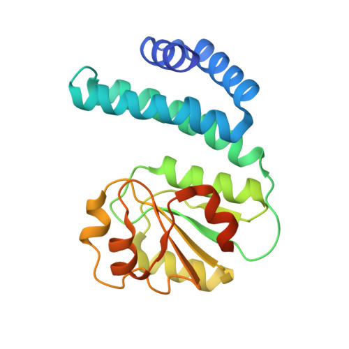
    CarH is a coenzyme B 12 -dependent photoreceptor involved in regulating carotenoid biosynthesis. How light-triggered cleavage of the B 12 Co-C bond culminates in CarH tetramer dissociation to initiate transcription remains unclear. Here, a series of crystal structures of the CarH B 12 -binding domain after illumination suggest formation of unforeseen intermediate states prior to tetramer dissociation. Unexpectedly, in the absence of oxygen, Co-C bond cleavage is followed by reorientation of the corrin ring and a switch from a lower to upper histidine-Co ligation, corresponding to a pentacoordinate state. Under aerobic conditions, rapid flash-cooling of crystals prior to deterioration upon illumination confirm a similar B 12 -ligand switch occurs. Removal of the upper His-ligating residue prevents monomer formation upon illumination. Combined with detailed solution spectroscopy and computational studies, these data demonstrate the CarH photoresponse integrates B 12 photo- and redox-chemistry to drive large-scale conformational changes through stepwise Co-ligation changes.


  • Organizational Affiliation
    • Manchester Institute of Biotechnology, Department of Chemistry, University of Manchester, Manchester, UK.

Macromolecule Content 

  • Total Structure Weight: 98.09 kDa 
  • Atom Count: 6,734 
  • Modeled Residue Count: 775 
  • Deposited Residue Count: 860 
  • Unique protein chains: 1

Macromolecules

Find similar proteins by:|  3D Structure
Entity ID: 1
MoleculeChains  Sequence LengthOrganismDetailsImage
Probable transcriptional regulator
A, B, C, D
215Thermus thermophilus HB27Mutation(s): 1 
Gene Names: TT_P0056
UniProt
Find proteins for Q746J7 (Thermus thermophilus (strain ATCC BAA-163 / DSM 7039 / HB27))
Explore Q746J7 
Go to UniProtKB:  Q746J7
Entity Groups
Sequence Clusters30% Identity50% Identity70% Identity90% Identity95% Identity100% Identity
UniProt GroupQ746J7
Sequence Annotations
Expand
Reference Sequence

Experimental Data & Validation

Experimental Data

  • Method: X-RAY DIFFRACTION
  • Resolution: 2.00 Å
  • R-Value Free:  0.218 (Depositor), 0.230 (DCC) 
  • R-Value Work:  0.181 (Depositor), 0.195 (DCC) 
  • R-Value Observed: 0.183 (Depositor) 
Space Group: P 21 21 21
Unit Cell:
Length ( Å )Angle ( ˚ )
a = 63.405α = 90
b = 70.664β = 90
c = 203.687γ = 90
Software Package:
Software NamePurpose
REFMACrefinement
xia2data reduction
DIALSdata scaling
PHASERphasing

Structure Validation

View Full Validation Report


Ligand Structure Quality Assessment 


Entry History 

& Funding Information

Deposition Data


Funding OrganizationLocationGrant Number
Engineering and Physical Sciences Research CouncilUnited KingdomEP/S030336/1

Revision History  (Full details and data files)

  • Version 1.0: 2023-08-16
    Type: Initial release
  • Version 1.1: 2023-08-23
    Changes: Refinement description
  • Version 1.2: 2023-08-30
    Changes: Database references