8C0J

Structure of AmiB enzymatic domain bound to the EnvC LytM domain


Experimental Data Snapshot

  • Method: X-RAY DIFFRACTION
  • Resolution: 3.38 Å
  • R-Value Free: 0.291 
  • R-Value Work: 0.249 

wwPDB Validation   3D Report Full Report


This is version 1.0 of the entry. See complete history


Literature

Activator-induced conformational changes regulate division-associated peptidoglycan amidases.

Cook, J.Baverstock, T.C.McAndrew, M.B.L.Roper, D.I.Stansfeld, P.J.Crow, A.

(2023) Proc Natl Acad Sci U S A 120: e2302580120-e2302580120

  • DOI: https://doi.org/10.1073/pnas.2302580120
  • Primary Citation of Related Structures:  
    8C0J, 8C2O

  • PubMed Abstract: 

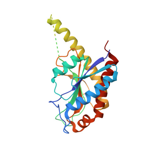
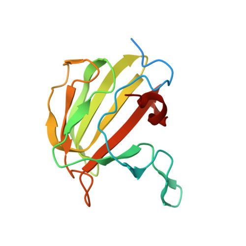
    AmiA and AmiB are peptidoglycan-hydrolyzing enzymes from Escherichia coli  that are required to break the peptidoglycan layer during bacterial cell division and maintain integrity of the cell envelope. In vivo, the activity of AmiA and AmiB is tightly controlled through their interactions with the membrane-bound FtsEX-EnvC complex. Activation of AmiA and AmiB requires access to a groove in the amidase-activating LytM domain of EnvC which is gated by ATP-driven conformational changes in FtsEX-EnvC complex. Here, we present a high-resolution structure of the isolated AmiA protein, confirming that it is autoinhibited in the same manner as AmiB and AmiC, and a complex of the AmiB enzymatic domain bound to the activating EnvC LytM domain. In isolation, the active site of AmiA is blocked by an autoinhibitory helix that binds directly to the catalytic zinc and fills the volume expected to accommodate peptidoglycan binding. In the complex, binding of the EnvC LytM domain induces a conformational change that displaces the amidase autoinhibitory helix and reorganizes the active site for activity. Our structures, together with complementary mutagenesis work, defines the conformational changes required to activate AmiA and/or AmiB through their interaction with their cognate activator EnvC.


  • Organizational Affiliation

    School of Life Sciences, University of Warwick, Coventry CV4 7AL, United Kingdom.


Macromolecules
Find similar proteins by:  (by identity cutoff)  |  3D Structure
Entity ID: 1
MoleculeChains Sequence LengthOrganismDetailsImage
N-acetylmuramoyl-L-alanine amidase
A, C
234Citrobacter rodentiumMutation(s): 0 
Gene Names: amiBE2R62_12775
EC: 3.5.1.28
UniProt
Find proteins for A0A482PQR2 (Citrobacter rodentium)
Explore A0A482PQR2 
Go to UniProtKB:  A0A482PQR2
Entity Groups  
Sequence Clusters30% Identity50% Identity70% Identity90% Identity95% Identity100% Identity
UniProt GroupA0A482PQR2
Sequence Annotations
Expand
  • Reference Sequence
Find similar proteins by:  (by identity cutoff)  |  3D Structure
Entity ID: 2
MoleculeChains Sequence LengthOrganismDetailsImage
Murein hydrolase activator EnvC149Citrobacter rodentiumMutation(s): 0 
Gene Names: envCE2R62_17750
UniProt
Find proteins for A0A482PID1 (Citrobacter rodentium)
Explore A0A482PID1 
Go to UniProtKB:  A0A482PID1
Entity Groups  
Sequence Clusters30% Identity50% Identity70% Identity90% Identity95% Identity100% Identity
UniProt GroupA0A482PID1
Sequence Annotations
Expand
  • Reference Sequence
Experimental Data & Validation

Experimental Data

  • Method: X-RAY DIFFRACTION
  • Resolution: 3.38 Å
  • R-Value Free: 0.291 
  • R-Value Work: 0.249 
  • Space Group: I 41 3 2
Unit Cell:
Length ( Å )Angle ( ˚ )
a = 237.363α = 90
b = 237.363β = 90
c = 237.363γ = 90
Software Package:
Software NamePurpose
REFMACrefinement
DIALSdata reduction
Aimlessdata scaling
PHASERphasing

Structure Validation

View Full Validation Report


Entry History & Funding Information

Deposition Data

  • Released Date: 2023-06-14 
  • Deposition Author(s): Crow, A.

Funding OrganizationLocationGrant Number
Biotechnology and Biological Sciences Research Council (BBSRC)United KingdomBB/V017101/1

Revision History  (Full details and data files)

  • Version 1.0: 2023-06-14
    Type: Initial release