8BZO

Cryo-EM structure of CDK2-CyclinA in complex with p27 from the SCFSKP2 E3 ligase Complex


Experimental Data Snapshot

  • Method: ELECTRON MICROSCOPY
  • Resolution: 3.50 Å
  • Aggregation State: PARTICLE 
  • Reconstruction Method: SINGLE PARTICLE 

wwPDB Validation   3D Report Full Report


This is version 1.1 of the entry. See complete history


Literature

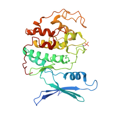
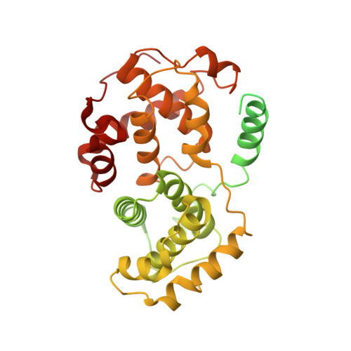
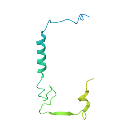
Cryo-EM structure of SKP1-SKP2-CKS1 in complex with CDK2-cyclin A-p27KIP1.

Rowland, R.J.Heath, R.Maskell, D.Thompson, R.F.Ranson, N.A.Blaza, J.N.Endicott, J.A.Noble, M.E.M.Salamina, M.

(2023) Sci Rep 13: 10718-10718

  • DOI: https://doi.org/10.1038/s41598-023-37609-9
  • Primary Citation of Related Structures:  
    8BYA, 8BYL, 8BZO

  • PubMed Abstract: 

    p27KIP1 (cyclin-dependent kinase inhibitor 1B, p27) is a member of the CIP/KIP family of CDK (cyclin dependent kinase) regulators that inhibit cell cycle CDKs. p27 phosphorylation by CDK1/2, signals its recruitment to the SCF SKP2 (S-phase kinase associated protein 1 (SKP1)-cullin-SKP2) E3 ubiquitin ligase complex for proteasomal degradation. The nature of p27 binding to SKP2 and CKS1 was revealed by the SKP1-SKP2-CKS1-p27 phosphopeptide crystal structure. Subsequently, a model for the hexameric CDK2-cyclin A-CKS1-p27-SKP1-SKP2 complex was proposed by overlaying an independently determined CDK2-cyclin A-p27 structure. Here we describe the experimentally determined structure of the isolated CDK2-cyclin A-CKS1-p27-SKP1-SKP2 complex at 3.4 Å global resolution using cryogenic electron microscopy. This structure supports previous analysis in which p27 was found to be structurally dynamic, transitioning from disordered to nascent secondary structure on target binding. We employed 3D variability analysis to further explore the conformational space of the hexameric complex and uncovered a previously unidentified hinge motion centred on CKS1. This flexibility gives rise to open and closed conformations of the hexameric complex that we propose may contribute to p27 regulation by facilitating recognition with SCF SKP2 . This 3D variability analysis further informed particle subtraction and local refinement approaches to enhance the local resolution of the complex.


  • Organizational Affiliation

    Translational and Clinical Research Institute, Newcastle University Centre for Cancer, Newcastle University, Paul O'Gorman Building, Framlington Place, Newcastle Upon Tyne, NE2 4HH, UK.


Macromolecules
Find similar proteins by:  (by identity cutoff)  |  3D Structure
Entity ID: 1
MoleculeChains Sequence LengthOrganismDetailsImage
Cyclin-dependent kinase 2297Homo sapiensMutation(s): 0 
Gene Names: CDK2CDKN2
EC: 2.7.11.22
UniProt & NIH Common Fund Data Resources
Find proteins for P24941 (Homo sapiens)
Explore P24941 
Go to UniProtKB:  P24941
PHAROS:  P24941
GTEx:  ENSG00000123374 
Entity Groups  
Sequence Clusters30% Identity50% Identity70% Identity90% Identity95% Identity100% Identity
UniProt GroupP24941
Sequence Annotations
Expand
  • Reference Sequence
Find similar proteins by:  (by identity cutoff)  |  3D Structure
Entity ID: 2
MoleculeChains Sequence LengthOrganismDetailsImage
Cyclin-A2432Homo sapiensMutation(s): 0 
Gene Names: CCNA2CCN1CCNA
UniProt & NIH Common Fund Data Resources
Find proteins for P20248 (Homo sapiens)
Explore P20248 
Go to UniProtKB:  P20248
PHAROS:  P20248
GTEx:  ENSG00000145386 
Entity Groups  
Sequence Clusters30% Identity50% Identity70% Identity90% Identity95% Identity100% Identity
UniProt GroupP20248
Sequence Annotations
Expand
  • Reference Sequence
Find similar proteins by:  (by identity cutoff)  |  3D Structure
Entity ID: 3
MoleculeChains Sequence LengthOrganismDetailsImage
Cyclin-dependent kinase inhibitor 1B158Homo sapiensMutation(s): 0 
Gene Names: p27 kip1
UniProt & NIH Common Fund Data Resources
Find proteins for P46527 (Homo sapiens)
Explore P46527 
Go to UniProtKB:  P46527
PHAROS:  P46527
GTEx:  ENSG00000111276 
Entity Groups  
Sequence Clusters30% Identity50% Identity70% Identity90% Identity95% Identity100% Identity
UniProt GroupP46527
Sequence Annotations
Expand
  • Reference Sequence
Small Molecules
Modified Residues  1 Unique
IDChains TypeFormula2D DiagramParent
TPO
Query on TPO
A
L-PEPTIDE LINKINGC4 H10 N O6 PTHR
Experimental Data & Validation

Experimental Data

  • Method: ELECTRON MICROSCOPY
  • Resolution: 3.50 Å
  • Aggregation State: PARTICLE 
  • Reconstruction Method: SINGLE PARTICLE 
EM Software:
TaskSoftware PackageVersion
MODEL REFINEMENTPHENIX
RECONSTRUCTIONcryoSPARC

Structure Validation

View Full Validation Report


Entry History & Funding Information

Deposition Data


Funding OrganizationLocationGrant Number
Medical Research Council (MRC, United Kingdom)United KingdomMR/N009738/1 and MR/V029142/1

Revision History  (Full details and data files)

  • Version 1.0: 2023-06-28
    Type: Initial release
  • Version 1.1: 2023-07-19
    Changes: Database references