Chromophore charge-state switching through copper-dependent homodimerisation of an engineered green fluorescent protein.
Ahmed, R.D., Vitsupakorn, D., Hartwell, K.D., Albalawi, K., Rizkallah, P.J., Watson, P.D., Jones, D.D.(2025) Chem Sci
- PubMed: 41142394 Search on PubMedSearch on PubMed Central
- DOI: https://doi.org/10.1039/d5sc06589e
- Primary Citation Related Structures:
8BXP, 8C1X - PubMed Abstract:
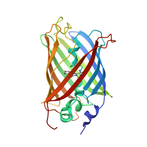
Here, we have linked one of the most common protein-protein interaction events, homodimerisation, to an essential trace metal, copper, through engineering green fluorescent protein. Mutation of H148 to cysteine promotes the neutral phenolic chromophore in the monomer that excites predominantly at ∼400 nm. Homodimerisation via a copper-dependent disulphide bridge switches the chromophore to the charged phenolate that excites at ∼490 nm. The result is a ∼30 fold increase in the fluorescence emission ratio. Homodimerisation kinetics are further improved by optimising the sfGFP homodimer interface, generating the variant termed GFP-diS2. Structures of the monomeric and dimeric GFP-diS2 suggest that charge switching is through peptide bond flipping and the formation of a buried organised water network around the chromophore that spans the interface region. Fusion to a leucine zipper protein dimerisation element greatly increased the GFP-diS2 association rate making it a more effective copper sensor in vitro and in vivo with Cu(i) instigating the signal change quicker and at lower ion concentrations than Cu(ii). Thus, GFP-diS2 provides the framework for generating a sensitive genetically encoded copper sensor and will eventually be adapted to monitor one of the most important protein-protein interactions in biology, homo-oligomerisation.
- School of Biosciences, Molecular Biosciences Division, Cardiff University Sir Martin Evans Building Cardiff CF10 3AX UK jonesdd@cardiff.ac.uk.
Organizational Affiliation: