8BT4

Ribonucleotide Reductase class Ie R2 from Mesoplasma florum, radical-lost ground state


Experimental Data Snapshot

  • Method: X-RAY DIFFRACTION
  • Resolution: 1.35 Å
  • R-Value Free: 0.164 
  • R-Value Work: 0.143 
  • R-Value Observed: 0.144 

wwPDB Validation   3D Report Full Report


This is version 1.0 of the entry. See complete history


Literature

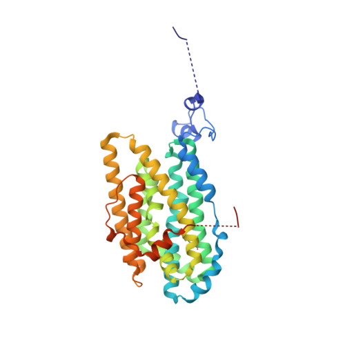
Structure of a ribonucleotide reductase R2 protein radical.

Lebrette, H.Srinivas, V.John, J.Aurelius, O.Kumar, R.Lundin, D.Brewster, A.S.Bhowmick, A.Sirohiwal, A.Kim, I.S.Gul, S.Pham, C.Sutherlin, K.D.Simon, P.Butryn, A.Aller, P.Orville, A.M.Fuller, F.D.Alonso-Mori, R.Batyuk, A.Sauter, N.K.Yachandra, V.K.Yano, J.Kaila, V.R.I.Sjoberg, B.M.Kern, J.Roos, K.Hogbom, M.

(2023) Science 382: 109-113

  • DOI: https://doi.org/10.1126/science.adh8160
  • Primary Citation of Related Structures:  
    8BT3, 8BT4

  • PubMed Abstract: 

    Aerobic ribonucleotide reductases (RNRs) initiate synthesis of DNA building blocks by generating a free radical within the R2 subunit; the radical is subsequently shuttled to the catalytic R1 subunit through proton-coupled electron transfer (PCET). We present a high-resolution room temperature structure of the class Ie R2 protein radical captured by x-ray free electron laser serial femtosecond crystallography. The structure reveals conformational reorganization to shield the radical and connect it to the translocation path, with structural changes propagating to the surface where the protein interacts with the catalytic R1 subunit. Restructuring of the hydrogen bond network, including a notably short O-O interaction of 2.41 angstroms, likely tunes and gates the radical during PCET. These structural results help explain radical handling and mobilization in RNR and have general implications for radical transfer in proteins.


  • Organizational Affiliation

    Department of Biochemistry and Biophysics, Stockholm University, Arrhenius Laboratories for Natural Sciences, Stockholm, Sweden.


Macromolecules
Find similar proteins by:  (by identity cutoff)  |  3D Structure
Entity ID: 1
MoleculeChains Sequence LengthOrganismDetailsImage
Ribonucleoside-diphosphate reductase
A, B
345Mesoplasma florum L1Mutation(s): 1 
Gene Names: Mfl530
EC: 1.17.4.1
UniProt
Find proteins for Q6F0T5 (Mesoplasma florum (strain ATCC 33453 / NBRC 100688 / NCTC 11704 / L1))
Explore Q6F0T5 
Go to UniProtKB:  Q6F0T5
Entity Groups  
Sequence Clusters30% Identity50% Identity70% Identity90% Identity95% Identity100% Identity
UniProt GroupQ6F0T5
Sequence Annotations
Expand
  • Reference Sequence
Small Molecules
Modified Residues  1 Unique
IDChains TypeFormula2D DiagramParent
DAH
Query on DAH
A, B
L-PEPTIDE LINKINGC9 H11 N O4PHE
Experimental Data & Validation

Experimental Data

  • Method: X-RAY DIFFRACTION
  • Resolution: 1.35 Å
  • R-Value Free: 0.164 
  • R-Value Work: 0.143 
  • R-Value Observed: 0.144 
  • Space Group: C 1 2 1
Unit Cell:
Length ( Å )Angle ( ˚ )
a = 175.503α = 90
b = 53.369β = 108.379
c = 79.03γ = 90
Software Package:
Software NamePurpose
XDSdata reduction
XDSdata scaling
PHENIXphasing
PHENIXrefinement
Cootmodel building

Structure Validation

View Full Validation Report


Entry History & Funding Information

Deposition Data


Funding OrganizationLocationGrant Number
Swedish Research CouncilSweden2021-03992
European Research Council (ERC)European UnionHIGH-GEAR 724394
Knut and Alice Wallenberg FoundationSweden2017.0275
Knut and Alice Wallenberg FoundationSweden2019.0436

Revision History  (Full details and data files)

  • Version 1.0: 2023-11-01
    Type: Initial release