8BPJ

X-ray structure of the adduct formed upon reaction of Lysozyme with [Ru2Cl(D-p-FPhF)(O2CCH3)3] (Structure 1)


Experimental Data Snapshot

  • Method: X-RAY DIFFRACTION
  • Resolution: 1.38 Å
  • R-Value Free: 0.231 
  • R-Value Work: 0.190 

wwPDB Validation   3D Report Full Report


Ligand Structure Quality Assessment 


This is version 1.1 of the entry. See complete history


Literature

Effect of Equatorial Ligand Substitution on the Reactivity with Proteins of Paddlewheel Diruthenium Complexes: Structural Studies.

Teran, A.Ferraro, G.Sanchez-Pelaez, A.E.Herrero, S.Merlino, A.

(2023) Inorg Chem 62: 670-674

  • DOI: https://doi.org/10.1021/acs.inorgchem.2c04103
  • Primary Citation of Related Structures:  
    8BPH, 8BPJ, 8BPU, 8BQM

  • PubMed Abstract: 

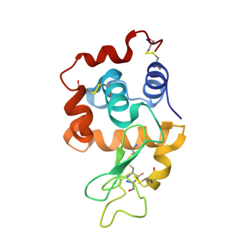
    The paddlewheel [Ru 2 Cl(O 2 CCH 3 ) 4 ] complex was previously reported to react with the model protein hen egg white lysozyme (HEWL), forming adducts with two diruthenium moieties bound to Asp101 and Asp119 side chains upon the release of one acetate. To study the effect of the equatorial ligands on the reactivity with proteins of diruthenium compounds, X-ray structures of the adducts formed when HEWL reacts with [Ru 2 Cl(D- p -FPhF)(O 2 CCH 3 ) 3 ] [D- p -FPhF = N , N '-bis(4-fluorophenyl)formamidinate] under different conditions were solved. [Ru 2 Cl(D- p -FPhF)(O 2 CCH 3 ) 3 ] is bonded through their equatorial positions to the Asp side chains. Protein binding occurs cis or trans to D- p -FPhF. Lys or Arg side chains or even main-chain carbonyl groups can coordinate to the diruthenium core at the axial site. Data help to understand the reactivity of paddlewheel diruthenium complexes with proteins, providing useful information for the design of new artificial diruthenium-containing metalloenzymes with potential applications in the fields of catalysis, biomedicine, and biotechnology.


  • Organizational Affiliation

    Departamento de Química Inorgánica, Facultad de Ciencias Químicas, Universidad Complutense de Madrid, Madrid E-28040, Spain.


Macromolecules
Find similar proteins by:  (by identity cutoff)  |  3D Structure
Entity ID: 1
MoleculeChains Sequence LengthOrganismDetailsImage
LysozymeA [auth AAA]129Gallus gallusMutation(s): 0 
UniProt
Find proteins for P00698 (Gallus gallus)
Explore P00698 
Go to UniProtKB:  P00698
Entity Groups  
Sequence Clusters30% Identity50% Identity70% Identity90% Identity95% Identity100% Identity
UniProt GroupP00698
Sequence Annotations
Expand
  • Reference Sequence
Small Molecules
Ligands 3 Unique
IDChains Name / Formula / InChI Key2D Diagram3D Interactions
TZ6 (Subject of Investigation/LOI)
Query on TZ6

Download Ideal Coordinates CCD File 
H [auth AAA],
I [auth AAA]
9,11-bis(4-fluorophenyl)-3,7-dimethyl-2,4,6,8-tetraoxa-9,11-diaza-1$l^{4},5$l^{4}-diruthenatricyclo[3.3.3.0^{1,5}]undecane
C17 H18 F2 N2 O4 Ru2
NOQXZAUOMFZMHS-UHFFFAOYSA-N
NO3
Query on NO3

Download Ideal Coordinates CCD File 
C [auth AAA],
D [auth AAA],
E [auth AAA],
F [auth AAA],
G [auth AAA]
NITRATE ION
N O3
NHNBFGGVMKEFGY-UHFFFAOYSA-N
NA
Query on NA

Download Ideal Coordinates CCD File 
B [auth AAA]SODIUM ION
Na
FKNQFGJONOIPTF-UHFFFAOYSA-N
Experimental Data & Validation

Experimental Data

  • Method: X-RAY DIFFRACTION
  • Resolution: 1.38 Å
  • R-Value Free: 0.231 
  • R-Value Work: 0.190 
  • Space Group: P 43 21 2
Unit Cell:
Length ( Å )Angle ( ˚ )
a = 77.48α = 90
b = 77.48β = 90
c = 37.27γ = 90
Software Package:
Software NamePurpose
REFMACrefinement
autoPROCdata reduction
autoPROCdata scaling
PHASERphasing

Structure Validation

View Full Validation Report


Ligand Structure Quality Assessment 


Entry History & Funding Information

Deposition Data


Funding OrganizationLocationGrant Number
Not funded--

Revision History  (Full details and data files)

  • Version 1.0: 2023-06-28
    Type: Initial release
  • Version 1.1: 2024-02-07
    Changes: Data collection, Refinement description