Effect of Equatorial Ligand Substitution on the Reactivity with Proteins of Paddlewheel Diruthenium Complexes: Structural Studies.
Teran, A., Ferraro, G., Sanchez-Pelaez, A.E., Herrero, S., Merlino, A.(2023) Inorg Chem 62: 670-674
- PubMed: 36597851
- DOI: https://doi.org/10.1021/acs.inorgchem.2c04103
- Primary Citation of Related Structures:
8BPH, 8BPJ, 8BPU, 8BQM - PubMed Abstract:
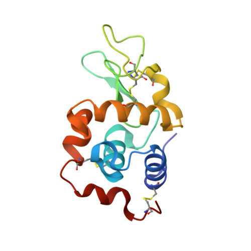
The paddlewheel [Ru 2 Cl(O 2 CCH 3 ) 4 ] complex was previously reported to react with the model protein hen egg white lysozyme (HEWL), forming adducts with two diruthenium moieties bound to Asp101 and Asp119 side chains upon the release of one acetate. To study the effect of the equatorial ligands on the reactivity with proteins of diruthenium compounds, X-ray structures of the adducts formed when HEWL reacts with [Ru 2 Cl(D- p -FPhF)(O 2 CCH 3 ) 3 ] [D- p -FPhF = N , N '-bis(4-fluorophenyl)formamidinate] under different conditions were solved. [Ru 2 Cl(D- p -FPhF)(O 2 CCH 3 ) 3 ] is bonded through their equatorial positions to the Asp side chains. Protein binding occurs cis or trans to D- p -FPhF. Lys or Arg side chains or even main-chain carbonyl groups can coordinate to the diruthenium core at the axial site. Data help to understand the reactivity of paddlewheel diruthenium complexes with proteins, providing useful information for the design of new artificial diruthenium-containing metalloenzymes with potential applications in the fields of catalysis, biomedicine, and biotechnology.
- Departamento de Química Inorgánica, Facultad de Ciencias Químicas, Universidad Complutense de Madrid, Madrid E-28040, Spain.
Organizational Affiliation:


