8BB7

Crystal structure of Mouse Plexin-B1 (20-535) in complex with VHH15


Experimental Data Snapshot

  • Method: X-RAY DIFFRACTION
  • Resolution: 2.95 Å
  • R-Value Free: 0.229 
  • R-Value Work: 0.203 
  • R-Value Observed: 0.204 

wwPDB Validation   3D Report Full Report


Ligand Structure Quality Assessment 


This is version 1.2 of the entry. See complete history


Literature

Nanobody inhibitors of Plexin-B1 identify allostery in plexin-semaphorin interactions and signaling.

Cowan, R.Trokter, M.Oleksy, A.Fedorova, M.Sawmynaden, K.Worzfeld, T.Offermanns, S.Matthews, D.Carr, M.D.Hall, G.

(2023) J Biol Chem 299: 104740-104740

  • DOI: https://doi.org/10.1016/j.jbc.2023.104740
  • Primary Citation of Related Structures:  
    8B3K, 8BB7, 8BF4

  • PubMed Abstract: 

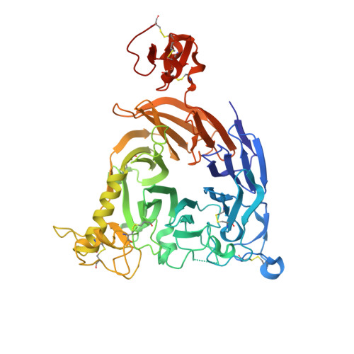
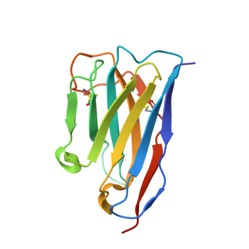
    Plexin-B1 is a receptor for the cell surface semaphorin, Sema4D. This signaling system has been implicated in a variety of human diseases, including cancer, multiple sclerosis and osteoporosis. While inhibitors of the Plexin-B1:Sema4D interaction have been previously reported, understanding their mechanism has been hindered by an incomplete structural view of Plexin-B1. In this study, we have raised and characterized a pair of nanobodies that are specific for mouse Plexin-B1 and which inhibit the binding of Sema4D to mouse Plexin-B1 and its biological activity. Structural studies of these nanobodies reveal that they inhibit the binding of Sema4D in an allosteric manner, binding to epitopes not previously reported. In addition, we report the first unbound structure of human Plexin-B1, which reveals that Plexin-B1 undergoes a conformational change on Sema4D binding. These changes mirror those seen upon binding of allosteric peptide modulators, which suggests a new model for understanding Plexin-B1 signaling and provides a potential innovative route for therapeutic modulation of Plexin-B1.


  • Organizational Affiliation

    Department of Molecular and Cell Biology, Leicester Institute of Structural and Chemical Biology, University of Leicester, Leicester, UK.


Macromolecules
Find similar proteins by:  (by identity cutoff)  |  3D Structure
Entity ID: 1
MoleculeChains Sequence LengthOrganismDetailsImage
Plexin-B1A [auth B],
C [auth A]
523Mus musculusMutation(s): 0 
Gene Names: Plxnb1Kiaa0407
UniProt
Find proteins for Q8CJH3 (Mus musculus)
Explore Q8CJH3 
Go to UniProtKB:  Q8CJH3
Entity Groups  
Sequence Clusters30% Identity50% Identity70% Identity90% Identity95% Identity100% Identity
UniProt GroupQ8CJH3
Sequence Annotations
Expand
  • Reference Sequence
Find similar proteins by:  (by identity cutoff)  |  3D Structure
Entity ID: 2
MoleculeChains Sequence LengthOrganismDetailsImage
VHH15 NanobodyB [auth D],
D [auth C]
130CamelidaeMutation(s): 0 
Entity Groups  
Sequence Clusters30% Identity50% Identity70% Identity90% Identity95% Identity100% Identity
Sequence Annotations
Expand
  • Reference Sequence
Experimental Data & Validation

Experimental Data

  • Method: X-RAY DIFFRACTION
  • Resolution: 2.95 Å
  • R-Value Free: 0.229 
  • R-Value Work: 0.203 
  • R-Value Observed: 0.204 
  • Space Group: P 1 21 1
Unit Cell:
Length ( Å )Angle ( ˚ )
a = 93.544α = 90
b = 108.988β = 90.37
c = 94.043γ = 90
Software Package:
Software NamePurpose
PHENIXrefinement
XDSdata reduction
Aimlessdata scaling
PHASERphasing
Cootmodel building

Structure Validation

View Full Validation Report


Ligand Structure Quality Assessment 


Entry History & Funding Information

Deposition Data


Funding OrganizationLocationGrant Number
Other privateUnited Kingdom--

Revision History  (Full details and data files)

  • Version 1.0: 2023-05-03
    Type: Initial release
  • Version 1.1: 2023-05-31
    Changes: Database references
  • Version 1.2: 2023-09-20
    Changes: Data collection, Refinement description