TMEM106B is a receptor mediating ACE2-independent SARS-CoV-2 cell entry.
Baggen, J., Jacquemyn, M., Persoons, L., Vanstreels, E., Pye, V.E., Wrobel, A.G., Calvaresi, V., Martin, S.R., Roustan, C., Cronin, N.B., Reading, E., Thibaut, H.J., Vercruysse, T., Maes, P., De Smet, F., Yee, A., Nivitchanyong, T., Roell, M., Franco-Hernandez, N., Rhinn, H., Mamchak, A.A., Ah Young-Chapon, M., Brown, E., Cherepanov, P., Daelemans, D.(2023) Cell 186: 3427
- PubMed: 37421949 Search on PubMedSearch on PubMed Central
- DOI: https://doi.org/10.1016/j.cell.2023.06.005
- Primary Citation Related Structures:
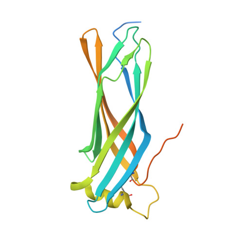
8B7D - PubMed Abstract:
SARS-CoV-2 is associated with broad tissue tropism, a characteristic often determined by the availability of entry receptors on host cells. Here, we show that TMEM106B, a lysosomal transmembrane protein, can serve as an alternative receptor for SARS-CoV-2 entry into angiotensin-converting enzyme 2 (ACE2)-negative cells. Spike substitution E484D increased TMEM106B binding, thereby enhancing TMEM106B-mediated entry. TMEM106B-specific monoclonal antibodies blocked SARS-CoV-2 infection, demonstrating a role of TMEM106B in viral entry. Using X-ray crystallography, cryogenic electron microscopy (cryo-EM), and hydrogen-deuterium exchange mass spectrometry (HDX-MS), we show that the luminal domain (LD) of TMEM106B engages the receptor-binding motif of SARS-CoV-2 spike. Finally, we show that TMEM106B promotes spike-mediated syncytium formation, suggesting a role of TMEM106B in viral fusion. Together, our findings identify an ACE2-independent SARS-CoV-2 infection mechanism that involves cooperative interactions with the receptors heparan sulfate and TMEM106B.
- KU Leuven Department of Microbiology, Immunology and Transplantation, Laboratory of Virology and Chemotherapy, Rega Institute, Leuven 3000, Belgium. Electronic address: jim.baggen@kuleuven.be.
Organizational Affiliation: