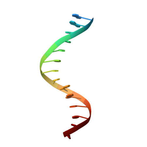
8ASC

Ku70/80 binds to the Ku-binding motif of PAXX


Experimental Data Snapshot

  • Method: X-RAY DIFFRACTION
  • Resolution: 2.95 Å
  • R-Value Free: 0.298 
  • R-Value Work: 0.252 
  • R-Value Observed: 0.255 

wwPDB Validation   3D Report Full Report


This is version 1.1 of the entry. See complete history


Literature

PAXX binding to the NHEJ machinery explains functional redundancy with XLF.

Seif-El-Dahan, M.Kefala-Stavridi, A.Frit, P.Hardwick, S.W.Chirgadze, D.Y.Maia De Oliviera, T.Britton, S.Barboule, N.Bossaert, M.Pandurangan, A.P.Meek, K.Blundell, T.L.Ropars, V.Calsou, P.Charbonnier, J.B.Chaplin, A.K.

(2023) Sci Adv 9: eadg2834-eadg2834

  • DOI: https://doi.org/10.1126/sciadv.adg2834
  • Primary Citation of Related Structures:  
    7ZWA, 7ZYG, 8ASC, 8BH3, 8BHV, 8BHY

  • PubMed Abstract: 

    Nonhomologous end joining is a critical mechanism that repairs DNA double-strand breaks in human cells. In this work, we address the structural and functional role of the accessory protein PAXX [paralog of x-ray repair cross-complementing protein 4 (XRCC4) and XRCC4-like factor (XLF)] in this mechanism. Here, we report high-resolution cryo-electron microscopy (cryo-EM) and x-ray crystallography structures of the PAXX C-terminal Ku-binding motif bound to Ku70/80 and cryo-EM structures of PAXX bound to two alternate DNA-dependent protein kinase (DNA-PK) end-bridging dimers, mediated by either Ku80 or XLF. We identify residues critical for the Ku70/PAXX interaction in vitro and in cells. We demonstrate that PAXX and XLF can bind simultaneously to the Ku heterodimer and act as structural bridges in alternate forms of DNA-PK dimers. Last, we show that engagement of both proteins provides a complementary advantage for DNA end synapsis and end joining in cells.


  • Organizational Affiliation

    Institute for Integrative Biology of the Cell (I2BC), Institute Joliot, CEA, CNRS, Université Paris-Saclay, 91198 Gif-sur-Yvette cedex, France.


Macromolecules

Find similar proteins by:  (by identity cutoff)  |  3D Structure
Entity ID: 1
MoleculeChains Sequence LengthOrganismDetailsImage
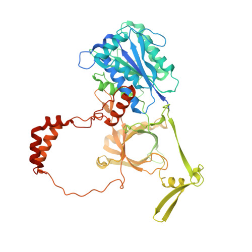
X-ray repair cross-complementing protein 6A,
E,
J [auth K],
N [auth O]
544Homo sapiensMutation(s): 0 
Gene Names: XRCC6G22P1
EC: 3.6.4 (PDB Primary Data), 4.2.99 (PDB Primary Data)
UniProt & NIH Common Fund Data Resources
Find proteins for P12956 (Homo sapiens)
Explore P12956 
Go to UniProtKB:  P12956
PHAROS:  P12956
GTEx:  ENSG00000196419 
Entity Groups  
Sequence Clusters30% Identity50% Identity70% Identity90% Identity95% Identity100% Identity
UniProt GroupP12956
Sequence Annotations
Expand
  • Reference Sequence
Find similar proteins by:  (by identity cutoff)  |  3D Structure
Entity ID: 2
MoleculeChains Sequence LengthOrganismDetailsImage
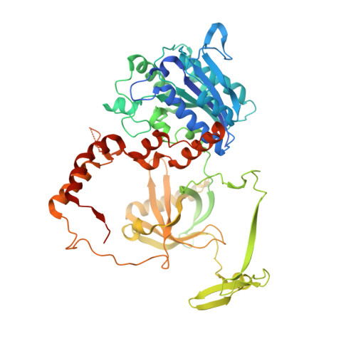
X-ray repair cross-complementing protein 5B,
F,
K [auth L],
O [auth P]
572Homo sapiensMutation(s): 0 
Gene Names: XRCC5G22P2
EC: 3.6.4
UniProt & NIH Common Fund Data Resources
Find proteins for P13010 (Homo sapiens)
Explore P13010 
Go to UniProtKB:  P13010
PHAROS:  P13010
GTEx:  ENSG00000079246 
Entity Groups  
Sequence Clusters30% Identity50% Identity70% Identity90% Identity95% Identity100% Identity
UniProt GroupP13010
Sequence Annotations
Expand
  • Reference Sequence
Find similar proteins by:  (by identity cutoff)  |  3D Structure
Entity ID: 5
MoleculeChains Sequence LengthOrganismDetailsImage
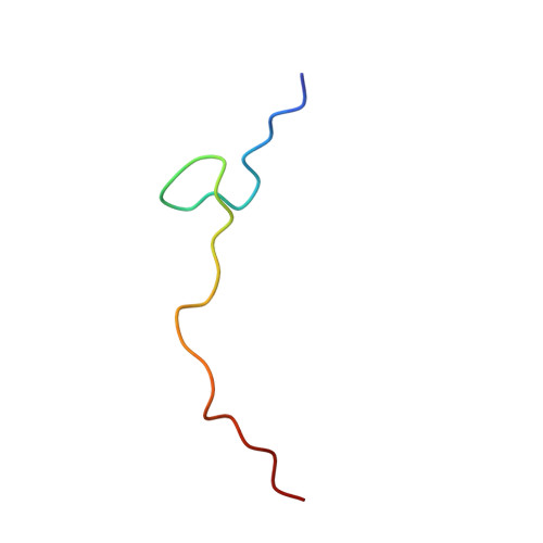
Protein PAXXI [auth J],
R [auth T]
28Homo sapiensMutation(s): 0 
UniProt & NIH Common Fund Data Resources
Find proteins for Q9BUH6 (Homo sapiens)
Explore Q9BUH6 
Go to UniProtKB:  Q9BUH6
GTEx:  ENSG00000148362 
Entity Groups  
Sequence Clusters30% Identity50% Identity70% Identity90% Identity95% Identity100% Identity
UniProt GroupQ9BUH6
Sequence Annotations
Expand
  • Reference Sequence
Find similar nucleic acids by:  (by identity cutoff)  |  3D Structure
Entity ID: 3
MoleculeChains LengthOrganismImage
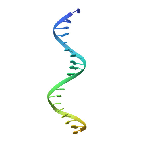
DNA (5'-D(P*CP*GP*GP*AP*TP*CP*GP*AP*GP*GP*GP*CP*CP*CP*GP*AP*TP*AP*T)-3')C,
G,
L [auth M],
P [auth Q]
30DNA molecule
Sequence Annotations
Expand
  • Reference Sequence

Find similar nucleic acids by:  Sequence   |   3D Structure  

Entity ID: 4
MoleculeChains LengthOrganismImage
DNA (5'-D(P*GP*GP*GP*CP*CP*CP*TP*CP*GP*AP*TP*CP*CP*G)-3')D,
H,
M [auth N],
Q [auth R]
15DNA molecule
Sequence Annotations
Expand
  • Reference Sequence
Experimental Data & Validation

Experimental Data

  • Method: X-RAY DIFFRACTION
  • Resolution: 2.95 Å
  • R-Value Free: 0.298 
  • R-Value Work: 0.252 
  • R-Value Observed: 0.255 
  • Space Group: P 1 21 1
Unit Cell:
Length ( Å )Angle ( ˚ )
a = 85.024α = 90
b = 428.57β = 90.01
c = 96.065γ = 90
Software Package:
Software NamePurpose
PHENIXrefinement
XDSdata reduction
STARANISOdata scaling
PHASERphasing

Structure Validation

View Full Validation Report


Entry History & Funding Information

Deposition Data


Funding OrganizationLocationGrant Number
French Alternative Energies and Atomic Energy Commission (CEA)FranceANR Break Dance

Revision History  (Full details and data files)

  • Version 1.0: 2023-06-21
    Type: Initial release
  • Version 1.1: 2024-02-07
    Changes: Data collection, Refinement description