7ZYG

CryoEM structure of Ku heterodimer bound to DNA, PAXX and XLF


Experimental Data Snapshot

  • Method: ELECTRON MICROSCOPY
  • Resolution: 2.68 Å
  • Aggregation State: PARTICLE 
  • Reconstruction Method: SINGLE PARTICLE 

wwPDB Validation   3D Report Full Report


This is version 2.0 of the entry. See complete history


Literature

PAXX binding to the NHEJ machinery explains functional redundancy with XLF.

Seif-El-Dahan, M.Kefala-Stavridi, A.Frit, P.Hardwick, S.W.Chirgadze, D.Y.Maia De Oliviera, T.Britton, S.Barboule, N.Bossaert, M.Pandurangan, A.P.Meek, K.Blundell, T.L.Ropars, V.Calsou, P.Charbonnier, J.B.Chaplin, A.K.

(2023) Sci Adv 9: eadg2834-eadg2834

  • DOI: https://doi.org/10.1126/sciadv.adg2834
  • Primary Citation of Related Structures:  
    7ZWA, 7ZYG, 8ASC, 8BH3, 8BHV, 8BHY

  • PubMed Abstract: 

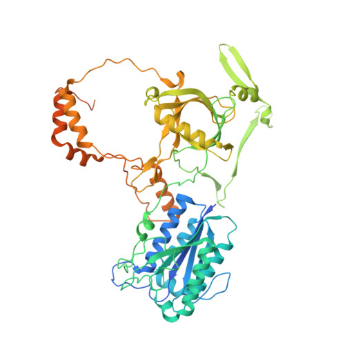
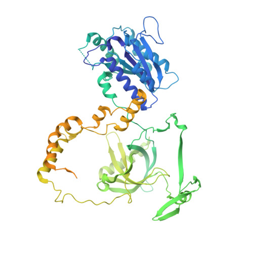
    Nonhomologous end joining is a critical mechanism that repairs DNA double-strand breaks in human cells. In this work, we address the structural and functional role of the accessory protein PAXX [paralog of x-ray repair cross-complementing protein 4 (XRCC4) and XRCC4-like factor (XLF)] in this mechanism. Here, we report high-resolution cryo-electron microscopy (cryo-EM) and x-ray crystallography structures of the PAXX C-terminal Ku-binding motif bound to Ku70/80 and cryo-EM structures of PAXX bound to two alternate DNA-dependent protein kinase (DNA-PK) end-bridging dimers, mediated by either Ku80 or XLF. We identify residues critical for the Ku70/PAXX interaction in vitro and in cells. We demonstrate that PAXX and XLF can bind simultaneously to the Ku heterodimer and act as structural bridges in alternate forms of DNA-PK dimers. Last, we show that engagement of both proteins provides a complementary advantage for DNA end synapsis and end joining in cells.


  • Organizational Affiliation

    Institute for Integrative Biology of the Cell (I2BC), Institute Joliot, CEA, CNRS, Université Paris-Saclay, 91198 Gif-sur-Yvette cedex, France.


Macromolecules

Find similar proteins by:  (by identity cutoff)  |  3D Structure
Entity ID: 1
MoleculeChains Sequence LengthOrganismDetailsImage
X-ray repair cross-complementing protein 6609Homo sapiensMutation(s): 0 
Gene Names: XRCC6G22P1
EC: 3.6.4 (PDB Primary Data), 4.2.99 (PDB Primary Data)
UniProt & NIH Common Fund Data Resources
Find proteins for P12956 (Homo sapiens)
Explore P12956 
Go to UniProtKB:  P12956
PHAROS:  P12956
GTEx:  ENSG00000196419 
Entity Groups  
Sequence Clusters30% Identity50% Identity70% Identity90% Identity95% Identity100% Identity
UniProt GroupP12956
Sequence Annotations
Expand
  • Reference Sequence
Find similar proteins by:  (by identity cutoff)  |  3D Structure
Entity ID: 2
MoleculeChains Sequence LengthOrganismDetailsImage
X-ray repair cross-complementing protein 5732Homo sapiensMutation(s): 0 
Gene Names: XRCC5G22P2
EC: 3.6.4
UniProt & NIH Common Fund Data Resources
Find proteins for P13010 (Homo sapiens)
Explore P13010 
Go to UniProtKB:  P13010
PHAROS:  P13010
GTEx:  ENSG00000079246 
Entity Groups  
Sequence Clusters30% Identity50% Identity70% Identity90% Identity95% Identity100% Identity
UniProt GroupP13010
Sequence Annotations
Expand
  • Reference Sequence
Find similar proteins by:  (by identity cutoff)  |  3D Structure
Entity ID: 3
MoleculeChains Sequence LengthOrganismDetailsImage
Protein PAXX204Homo sapiensMutation(s): 0 
Gene Names: PAXXC9orf142XLS
UniProt & NIH Common Fund Data Resources
Find proteins for Q9BUH6 (Homo sapiens)
Explore Q9BUH6 
Go to UniProtKB:  Q9BUH6
GTEx:  ENSG00000148362 
Entity Groups  
Sequence Clusters30% Identity50% Identity70% Identity90% Identity95% Identity100% Identity
UniProt GroupQ9BUH6
Sequence Annotations
Expand
  • Reference Sequence
Find similar proteins by:  (by identity cutoff)  |  3D Structure
Entity ID: 4
MoleculeChains Sequence LengthOrganismDetailsImage
Non-homologous end-joining factor 1D [auth F]299Homo sapiensMutation(s): 0 
Gene Names: NHEJ1XLF
UniProt & NIH Common Fund Data Resources
Find proteins for Q9H9Q4 (Homo sapiens)
Explore Q9H9Q4 
Go to UniProtKB:  Q9H9Q4
PHAROS:  Q9H9Q4
GTEx:  ENSG00000187736 
Entity Groups  
Sequence Clusters30% Identity50% Identity70% Identity90% Identity95% Identity100% Identity
UniProt GroupQ9H9Q4
Sequence Annotations
Expand
  • Reference Sequence

Find similar nucleic acids by:  Sequence   |   3D Structure  

Entity ID: 5
MoleculeChains LengthOrganismImage
DNAE [auth D]15Homo sapiens
Sequence Annotations
Expand
  • Reference Sequence

Find similar nucleic acids by:  Sequence   |   3D Structure  

Entity ID: 6
MoleculeChains LengthOrganismImage
DNAF [auth E]15Homo sapiens
Sequence Annotations
Expand
  • Reference Sequence
Small Molecules
Ligands 1 Unique
IDChains Name / Formula / InChI Key2D Diagram3D Interactions
PO4
Query on PO4

Download Ideal Coordinates CCD File 
G [auth B]PHOSPHATE ION
O4 P
NBIIXXVUZAFLBC-UHFFFAOYSA-K
Experimental Data & Validation

Experimental Data

  • Method: ELECTRON MICROSCOPY
  • Resolution: 2.68 Å
  • Aggregation State: PARTICLE 
  • Reconstruction Method: SINGLE PARTICLE 
EM Software:
TaskSoftware PackageVersion
RECONSTRUCTIONcryoSPARC
MODEL REFINEMENTPHENIX

Structure Validation

View Full Validation Report


Entry History & Funding Information

Deposition Data


Funding OrganizationLocationGrant Number
Wellcome TrustUnited Kingdom200814/Z/16/Z; 2016

Revision History  (Full details and data files)

  • Version 1.0: 2023-06-07
    Type: Initial release
  • Version 2.0: 2023-06-14
    Changes: Advisory, Atomic model, Data collection, Database references, Derived calculations, Polymer sequence, Source and taxonomy, Structure summary