8AIN | pdb_00008ain

MCUGI SAUNG complex


Experimental Data Snapshot

  • Method: X-RAY DIFFRACTION
  • Resolution: 2.70 Å
  • R-Value Free: 
    0.261 (Depositor), 0.264 (DCC) 
  • R-Value Work: 
    0.198 (Depositor), 0.202 (DCC) 

Starting Model: experimental
View more details

wwPDB Validation 3D Report Full Report

Validation slider image for 8AIN

This is version 1.3 of the entry. See complete history

Literature

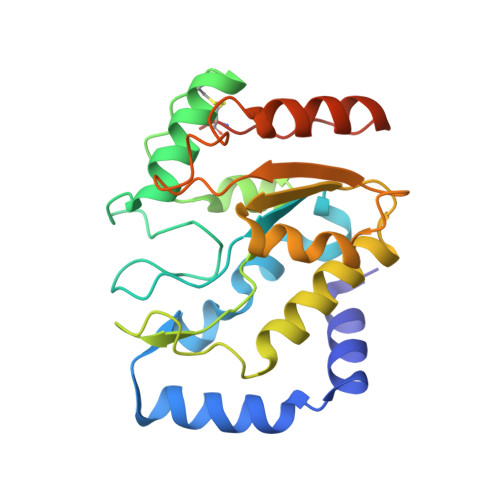
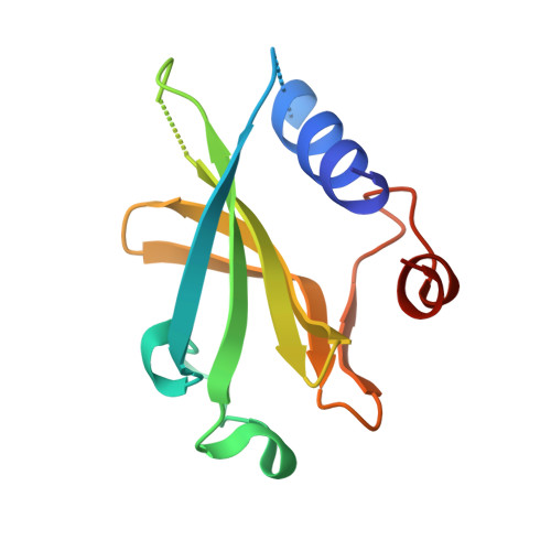
A Multimodal Approach towards Genomic Identification of Protein Inhibitors of Uracil-DNA Glycosylase.

Muselmani, W.Kashif-Khan, N.Bagneris, C.Santangelo, R.Williams, M.A.Savva, R.

(2023) Viruses 15

  • DOI: https://doi.org/10.3390/v15061348
  • Primary Citation Related Structures: 
    8AIL, 8AIM, 8AIN

  • PubMed Abstract: 

    DNA-mimicking proteins encoded by viruses can modulate processes such as innate cellular immunity. An example is Ung-family uracil-DNA glycosylase inhibition, which prevents Ung-mediated degradation via the stoichiometric protein blockade of the Ung DNA-binding cleft. This is significant where uracil-DNA is a key determinant in the replication and distribution of virus genomes. Unrelated protein folds support a common physicochemical spatial strategy for Ung inhibition, characterised by pronounced sequence plasticity within the diverse fold families. That, and the fact that relatively few template sequences are biochemically verified to encode Ung inhibitor proteins, presents a barrier to the straightforward identification of Ung inhibitors in genomic sequences. In this study, distant homologs of known Ung inhibitors were characterised via structural biology and structure prediction methods. A recombinant cellular survival assay and in vitro biochemical assay were used to screen distant variants and mutants to further explore tolerated sequence plasticity in motifs supporting Ung inhibition. The resulting validated sequence repertoire defines an expanded set of heuristic sequence and biophysical signatures shared by known Ung inhibitor proteins. A computational search of genome database sequences and the results of recombinant tests of selected output sequences obtained are presented here.


  • Organizational Affiliation
    • Institute of Structural and Molecular Biology, Department of Biological Sciences, Birkbeck, University of London, Malet Street, London WC1E 7HX, UK.

Macromolecule Content 

  • Total Structure Weight: 40.38 kDa 
  • Atom Count: 2,676 
  • Modeled Residue Count: 322 
  • Deposited Residue Count: 346 
  • Unique protein chains: 2

Macromolecules

Find similar proteins by:|  3D Structure
Entity ID: 1
MoleculeChains  Sequence LengthOrganismDetailsImage
Uracil-DNA glycosylase226Staphylococcus aureusMutation(s): 0 
Gene Names: 
EC: 3.2.2.27
UniProt
Find proteins for Q2G0J7 (Staphylococcus aureus (strain NCTC 8325 / PS 47))
Explore Q2G0J7 
Go to UniProtKB:  Q2G0J7
Entity Groups
Sequence Clusters30% Identity50% Identity70% Identity90% Identity95% Identity100% Identity
UniProt GroupQ2G0J7
Sequence Annotations
Expand
Reference Sequence
Find similar proteins by:|  3D Structure
Entity ID: 2
MoleculeChains  Sequence LengthOrganismDetailsImage
MCUGI120Macrococcoides caseolyticumMutation(s): 0 
UniProt
Find proteins for D4AH32 (Macrococcoides caseolyticum)
Explore D4AH32 
Go to UniProtKB:  D4AH32
Entity Groups
Sequence Clusters30% Identity50% Identity70% Identity90% Identity95% Identity100% Identity
UniProt GroupD4AH32
Sequence Annotations
Expand
Reference Sequence

Experimental Data & Validation

Experimental Data

  • Method: X-RAY DIFFRACTION
  • Resolution: 2.70 Å
  • R-Value Free:  0.261 (Depositor), 0.264 (DCC) 
  • R-Value Work:  0.198 (Depositor), 0.202 (DCC) 
Space Group: P 63 2 2
Unit Cell:
Length ( Å )Angle ( ˚ )
a = 91.522α = 90
b = 91.522β = 90
c = 158.62γ = 120
Software Package:
Software NamePurpose
REFMACrefinement
XDSdata reduction
Aimlessdata scaling
MOLREPphasing

Structure Validation

View Full Validation Report


Entry History 

& Funding Information

Deposition Data


Funding OrganizationLocationGrant Number
Birkbeck CollegeUnited Kingdom--

Revision History  (Full details and data files)

  • Version 1.0: 2023-06-21
    Type: Initial release
  • Version 1.1: 2023-07-12
    Changes: Database references
  • Version 1.2: 2024-02-07
    Changes: Data collection, Refinement description
  • Version 1.3: 2024-10-16
    Changes: Structure summary