8A5V

Crystal structure of the human phosposerine aminotransferase (PSAT)


Experimental Data Snapshot

  • Method: X-RAY DIFFRACTION
  • Resolution: 2.46 Å
  • R-Value Free: 0.210 
  • R-Value Work: 0.179 
  • R-Value Observed: 0.180 

wwPDB Validation   3D Report Full Report


Ligand Structure Quality Assessment 


This is version 1.2 of the entry. See complete history


Literature

L-serine biosynthesis in the human central nervous system: Structure and function of phosphoserine aminotransferase.

Marchesani, F.Zangelmi, E.Murtas, G.Costanzi, E.Ullah, R.Peracchi, A.Bruno, S.Pollegioni, L.Mozzarelli, A.Storici, P.Campanini, B.

(2023) Protein Sci 32: e4609-e4609

  • DOI: https://doi.org/10.1002/pro.4609
  • Primary Citation of Related Structures:  
    8A5V, 8A5W

  • PubMed Abstract: 

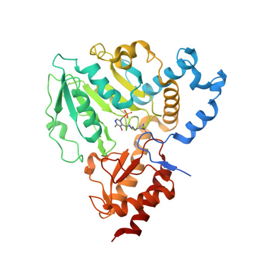
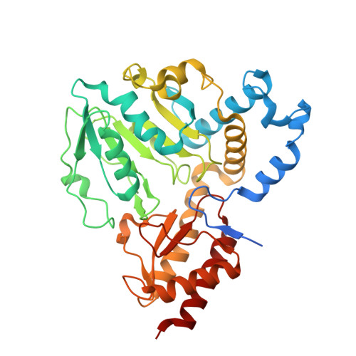
    Organisms from all kingdoms of life synthesize L-serine (L-Ser) from 3-phosphoglycerate through the phosphorylated pathway, a three-step diversion of glycolysis. Phosphoserine aminotransferase (PSAT) catalyzes the intermediate step, the pyridoxal 5'-phosphate-dependent transamination of 3-phosphohydroxypyruvate and L-glutamate to O-phosphoserine (OPS) and α-ketoglutarate. PSAT is particularly relevant in the central nervous system of mammals because L-Ser is the metabolic precursor of D-serine, cysteine, phospholipids, and nucleotides. Several mutations in the human psat gene have been linked to serine deficiency disorders, characterized by severe neurological symptoms. Furthermore, PSAT is overexpressed in many tumors and this overexpression has been associated with poor clinical outcomes. Here, we report the detailed functional and structural characterization of the recombinant human PSAT. The reaction catalyzed by PSAT is reversible, with an equilibrium constant of about 10, and the enzyme is very efficient, with a k cat /K m of 5.9 × 10 6  M -1  s -1 , thus contributing in driving the pathway towards the products despite the extremely unfavorable first step catalyzed by 3-phosphoglycerate dehydrogenase. The 3D X-ray crystal structure of PSAT was solved in the substrate-free as well as in the OPS-bound forms. Both structures contain eight protein molecules in the asymmetric unit, arranged in four dimers, with a bound cofactor in each subunit. In the substrate-free form, the active site of PSAT contains a sulfate ion that, in the substrate-bound form, is replaced by the phosphate group of OPS. Interestingly, fast crystal soaking used to produce the substrate-bound form allowed the trapping of different intermediates along the catalytic cycle.


  • Organizational Affiliation

    Department of Food and Drug, University of Parma, Parma, Italy.


Macromolecules
Find similar proteins by:  (by identity cutoff)  |  3D Structure
Entity ID: 1
MoleculeChains Sequence LengthOrganismDetailsImage
Phosphoserine aminotransferase393Homo sapiensMutation(s): 0 
Gene Names: PSAT1PSA
EC: 2.6.1.52
UniProt & NIH Common Fund Data Resources
Find proteins for Q9Y617 (Homo sapiens)
Explore Q9Y617 
Go to UniProtKB:  Q9Y617
PHAROS:  Q9Y617
GTEx:  ENSG00000135069 
Entity Groups  
Sequence Clusters30% Identity50% Identity70% Identity90% Identity95% Identity100% Identity
UniProt GroupQ9Y617
Sequence Annotations
Expand
  • Reference Sequence
Find similar proteins by:  (by identity cutoff)  |  3D Structure
Entity ID: 2
MoleculeChains Sequence LengthOrganismDetailsImage
Phosphoserine aminotransferaseD [auth B],
G
393Homo sapiensMutation(s): 0 
Gene Names: PSAT1PSA
EC: 2.6.1.52
UniProt & NIH Common Fund Data Resources
Find proteins for Q9Y617 (Homo sapiens)
Explore Q9Y617 
Go to UniProtKB:  Q9Y617
PHAROS:  Q9Y617
GTEx:  ENSG00000135069 
Entity Groups  
Sequence Clusters30% Identity50% Identity70% Identity90% Identity95% Identity100% Identity
UniProt GroupQ9Y617
Sequence Annotations
Expand
  • Reference Sequence
Small Molecules
Ligands 2 Unique
IDChains Name / Formula / InChI Key2D Diagram3D Interactions
PMP (Subject of Investigation/LOI)
Query on PMP

Download Ideal Coordinates CCD File 
L [auth B],
P [auth G]
4'-DEOXY-4'-AMINOPYRIDOXAL-5'-PHOSPHATE
C8 H13 N2 O5 P
ZMJGSOSNSPKHNH-UHFFFAOYSA-N
SO4
Query on SO4

Download Ideal Coordinates CCD File 
I [auth D]
J [auth E]
K [auth C]
M [auth B]
N [auth F]
I [auth D],
J [auth E],
K [auth C],
M [auth B],
N [auth F],
O [auth A],
Q [auth G],
R [auth H]
SULFATE ION
O4 S
QAOWNCQODCNURD-UHFFFAOYSA-L
Modified Residues  1 Unique
IDChains TypeFormula2D DiagramParent
LLP
Query on LLP
A [auth D]
B [auth E]
C
E [auth F]
F [auth A]
A [auth D],
B [auth E],
C,
E [auth F],
F [auth A],
H
L-PEPTIDE LINKINGC14 H22 N3 O7 PLYS
Experimental Data & Validation

Experimental Data

  • Method: X-RAY DIFFRACTION
  • Resolution: 2.46 Å
  • R-Value Free: 0.210 
  • R-Value Work: 0.179 
  • R-Value Observed: 0.180 
  • Space Group: P 1 21 1
Unit Cell:
Length ( Å )Angle ( ˚ )
a = 65.87α = 90
b = 206.268β = 98.004
c = 135.539γ = 90
Software Package:
Software NamePurpose
PHENIXrefinement
PHENIXrefinement
autoPROCdata reduction
STARANISOdata scaling
PHASERphasing

Structure Validation

View Full Validation Report


Ligand Structure Quality Assessment 


Entry History & Funding Information

Deposition Data


Funding OrganizationLocationGrant Number
Ministero dell Universita e della RicercaItaly2017H4J3A

Revision History  (Full details and data files)

  • Version 1.0: 2023-03-29
    Type: Initial release
  • Version 1.1: 2023-04-12
    Changes: Database references
  • Version 1.2: 2024-02-07
    Changes: Data collection, Refinement description