7ZOO

Carbohydrate binding domain CBM92-B from a multi-catalytic glucanase-chitinase from Chitinophaga pinensis DSM 2588 in complex with gentiobiose


Experimental Data Snapshot

  • Method: X-RAY DIFFRACTION
  • Resolution: 1.84 Å
  • R-Value Free: 0.216 
  • R-Value Work: 0.191 
  • R-Value Observed: 0.193 

wwPDB Validation   3D Report Full Report


Ligand Structure Quality Assessment 


This is version 2.2 of the entry. See complete history


Literature

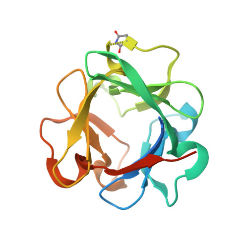
Structural and biochemical analysis of family 92 carbohydrate-binding modules uncovers multivalent binding to beta-glucans.

Hao, M.S.Mazurkewich, S.Li, H.Kvammen, A.Saha, S.Koskela, S.Inman, A.R.Nakajima, M.Tanaka, N.Nakai, H.Branden, G.Bulone, V.Larsbrink, J.McKee, L.S.

(2024) Nat Commun 15: 3429-3429

  • DOI: https://doi.org/10.1038/s41467-024-47584-y
  • Primary Citation of Related Structures:  
    7ZOH, 7ZOI, 7ZON, 7ZOO, 7ZOP

  • PubMed Abstract: 

    Carbohydrate-binding modules (CBMs) are non-catalytic proteins found appended to carbohydrate-active enzymes. Soil and marine bacteria secrete such enzymes to scavenge nutrition, and they often use CBMs to improve reaction rates and retention of released sugars. Here we present a structural and functional analysis of the recently established CBM family 92. All proteins analysed bind preferentially to β-1,6-glucans. This contrasts with the diversity of predicted substrates among the enzymes attached to CBM92 domains. We present crystal structures for two proteins, and confirm by mutagenesis that tryptophan residues permit ligand binding at three distinct functional binding sites on each protein. Multivalent CBM families are uncommon, so the establishment and structural characterisation of CBM92 enriches the classification database and will facilitate functional prediction in future projects. We propose that CBM92 proteins may cross-link polysaccharides in nature, and might have use in novel strategies for enzyme immobilisation.


  • Organizational Affiliation

    Division of Glycoscience, Department of Chemistry, KTH Royal Institute of Technology, AlbaNova University Centre, 106 91, Stockholm, Sweden.


Macromolecules
Find similar proteins by:  (by identity cutoff)  |  3D Structure
Entity ID: 1
MoleculeChains Sequence LengthOrganismDetailsImage
Glycoside hydrolase family 18
A, B
147Chitinophaga pinensis DSM 2588Mutation(s): 0 
Gene Names: Cpin_2580
UniProt
Find proteins for A0A979GQH9 (Chitinophaga pinensis (strain ATCC 43595 / DSM 2588 / LMG 13176 / NBRC 15968 / NCIMB 11800 / UQM 2034))
Explore A0A979GQH9 
Go to UniProtKB:  A0A979GQH9
Entity Groups  
Sequence Clusters30% Identity50% Identity70% Identity90% Identity95% Identity100% Identity
UniProt GroupA0A979GQH9
Sequence Annotations
Expand
  • Reference Sequence
Small Molecules
Experimental Data & Validation

Experimental Data

  • Method: X-RAY DIFFRACTION
  • Resolution: 1.84 Å
  • R-Value Free: 0.216 
  • R-Value Work: 0.191 
  • R-Value Observed: 0.193 
  • Space Group: C 1 2 1
Unit Cell:
Length ( Å )Angle ( ˚ )
a = 44.621α = 90
b = 111.407β = 98.575
c = 70.536γ = 90
Software Package:
Software NamePurpose
XDSdata reduction
XDSdata scaling
PHASERphasing
PHENIXrefinement

Structure Validation

View Full Validation Report


Ligand Structure Quality Assessment 


Entry History & Funding Information

Deposition Data


Funding OrganizationLocationGrant Number
Swedish Research CouncilSweden2017-04906
Knut and Alice Wallenberg FoundationSwedenWWSC

Revision History  (Full details and data files)

  • Version 1.0: 2023-05-10
    Type: Initial release
  • Version 2.0: 2023-11-15
    Changes: Atomic model, Data collection, Derived calculations
  • Version 2.1: 2024-02-07
    Changes: Refinement description
  • Version 2.2: 2024-05-08
    Changes: Database references, Derived calculations, Source and taxonomy, Structure summary