7ZMU

14-3-3s binding to non-natural peptide 2d


Experimental Data Snapshot

  • Method: X-RAY DIFFRACTION
  • Resolution: 1.60 Å
  • R-Value Free: 0.294 
  • R-Value Work: 0.254 
  • R-Value Observed: 0.256 

wwPDB Validation   3D Report Full Report


This is version 2.1 of the entry. See complete history


Literature

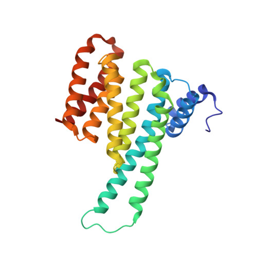
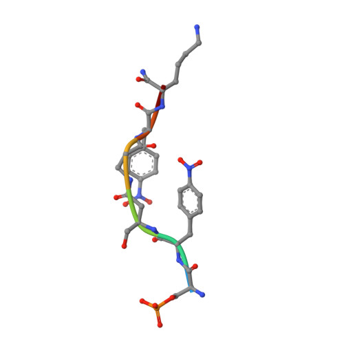
Functional mapping of the 14-3-3 hub protein as a guide to design 14-3-3 molecular glues.

Somsen, B.A.Craenmehr, F.W.B.Liu, W.W.Koops, A.A.Pennings, M.A.M.Visser, E.J.Ottmann, C.Cossar, P.J.Brunsveld, L.

(2022) Chem Sci 13: 13122-13131

  • DOI: https://doi.org/10.1039/d2sc04662h
  • Primary Citation of Related Structures:  
    7ZMU, 7ZMW

  • PubMed Abstract: 

    Molecular glues represent an evolution in drug discovery, however, targeted stabilization of protein complexes remains challenging, owing to a paucity of drug design rules. The functional mapping of hotspots has been critical to protein-protein interaction (PPI) inhibitor research, however, the orthogonal approach to stabilize PPIs has not exploited this information. Utilizing the hub protein 14-3-3 as a case study we demonstrate that functional mapping of hotspots provides a triage map for 14-3-3 molecular glue development. Truncation and mutation studies allowed deconvoluting the energetic contributions of sidechain and backbone interactions of a 14-3-3-binding non-natural peptide. Three central 14-3-3 hotspots were identified and their thermodynamic characteristics profiled. In addition to the phospho-binding pocket; (i) Asn226, (ii) Lys122 and (iii) the hydrophobic patch formed by Leu218, Ile219 and Leu222 were critical for protein complex formation. Exploiting this hotspot information allowed a peptide-based molecular glue that elicits high cooperativity ( α = 36) and selectively stabilizes the 14-3-3/ChREBP PPI to be uniquely developed.


  • Organizational Affiliation

    Laboratory of Chemical Biology, Department of Biomedical Engineering and Institute for Complex Molecular Systems, Eindhoven University of Technology P.O. Box 513 Eindhoven 5600 MB The Netherlands p.cossar@tue.nl l.brunsveld@tue.nl.


Macromolecules
Find similar proteins by:  (by identity cutoff)  |  3D Structure
Entity ID: 1
MoleculeChains Sequence LengthOrganismDetailsImage
14-3-3 protein sigma236Homo sapiensMutation(s): 0 
Gene Names: SFNHME1
UniProt & NIH Common Fund Data Resources
Find proteins for P31947 (Homo sapiens)
Explore P31947 
Go to UniProtKB:  P31947
PHAROS:  P31947
GTEx:  ENSG00000175793 
Entity Groups  
Sequence Clusters30% Identity50% Identity70% Identity90% Identity95% Identity100% Identity
UniProt GroupP31947
Sequence Annotations
Expand
  • Reference Sequence

Find similar proteins by:  Sequence   |   3D Structure  

Entity ID: 2
MoleculeChains Sequence LengthOrganismDetailsImage
non-natural peptide 27synthetic constructMutation(s): 0 
Sequence Annotations
Expand
  • Reference Sequence
Small Molecules
Modified Residues  2 Unique
IDChains TypeFormula2D DiagramParent
PPN
Query on PPN
B
L-PEPTIDE LINKINGC9 H10 N2 O4PHE
SEP
Query on SEP
B
L-PEPTIDE LINKINGC3 H8 N O6 PSER
Experimental Data & Validation

Experimental Data

  • Method: X-RAY DIFFRACTION
  • Resolution: 1.60 Å
  • R-Value Free: 0.294 
  • R-Value Work: 0.254 
  • R-Value Observed: 0.256 
  • Space Group: C 2 2 21
Unit Cell:
Length ( Å )Angle ( ˚ )
a = 82.278α = 90
b = 112.007β = 90
c = 62.68γ = 90
Software Package:
Software NamePurpose
PHENIXrefinement
PDB_EXTRACTdata extraction
Cootmodel building
Aimlessdata scaling
XDSdata reduction
MOLREPphasing

Structure Validation

View Full Validation Report


Entry History & Funding Information

Deposition Data


Funding OrganizationLocationGrant Number
Netherlands Organisation for Scientific Research (NWO)Netherlands711.017.014

Revision History  (Full details and data files)

  • Version 1.0: 2022-11-23
    Type: Initial release
  • Version 1.1: 2022-12-07
    Changes: Database references
  • Version 2.0: 2023-11-15
    Changes: Atomic model, Data collection
  • Version 2.1: 2024-01-31
    Changes: Refinement description