7Z9M

E.coli gyrase holocomplex with 217 bp DNA and Albi-1 (site AA)


Experimental Data Snapshot

  • Method: ELECTRON MICROSCOPY
  • Resolution: 3.30 Å
  • Aggregation State: PARTICLE 
  • Reconstruction Method: SINGLE PARTICLE 

wwPDB Validation   3D Report Full Report


This is version 1.1 of the entry. See complete history

Re-refinement Note

This entry reflects an alternative modeling of the original data in: 7Z9C 7Z9G


Literature

Molecular mechanism of topoisomerase poisoning by the peptide antibiotic albicidin.

Michalczyk, E.Hommernick, K.Behroz, I.Kulike, M.Pakosz-Stepien, Z.Mazurek, L.Seidel, M.Kunert, M.Santos, K.von Moeller, H.Loll, B.Weston, J.B.Mainz, A.Heddle, J.G.Sussmuth, R.D.Ghilarov, D.

(2023) Nat Catal 6: 52-67

  • DOI: https://doi.org/10.1038/s41929-022-00904-1
  • Primary Citation of Related Structures:  
    7Z9C, 7Z9G, 7Z9K, 7Z9M

  • PubMed Abstract: 

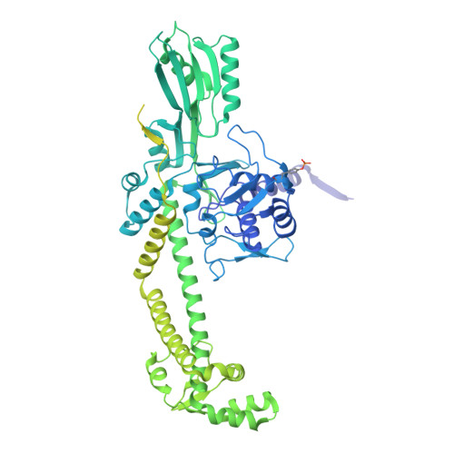
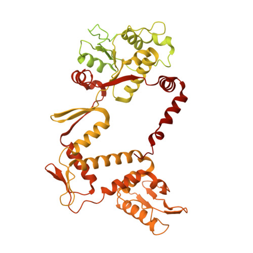
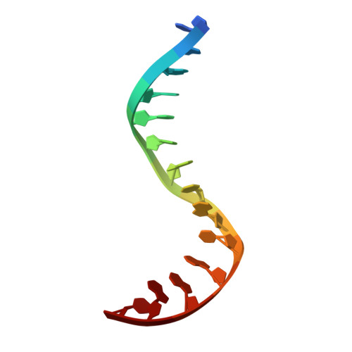
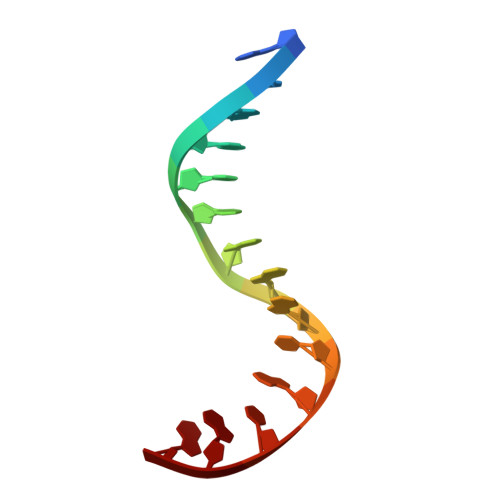
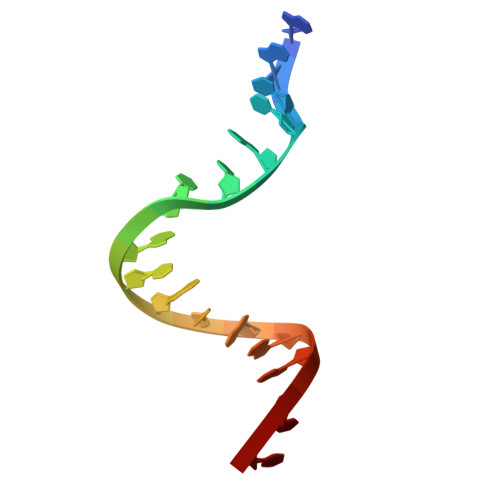
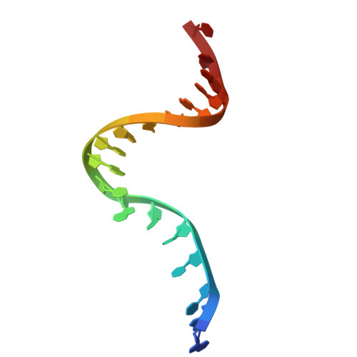
    The peptide antibiotic albicidin is a DNA topoisomerase inhibitor with low-nanomolar bactericidal activity towards fluoroquinolone-resistant Gram-negative pathogens. However, its mode of action is poorly understood. We determined a 2.6 Å resolution cryoelectron microscopy structure of a ternary complex between Escherichia coli topoisomerase DNA gyrase, a 217 bp double-stranded DNA fragment and albicidin. Albicidin employs a dual binding mechanism where one end of the molecule obstructs the crucial gyrase dimer interface, while the other intercalates between the fragments of cleaved DNA substrate. Thus, albicidin efficiently locks DNA gyrase, preventing it from religating DNA and completing its catalytic cycle. Two additional structures of this trapped state were determined using synthetic albicidin analogues that demonstrate improved solubility, and activity against a range of gyrase variants and E. coli topoisomerase IV. The extraordinary promiscuity of the DNA-intercalating region of albicidins and their excellent performance against fluoroquinolone-resistant bacteria holds great promise for the development of last-resort antibiotics.


  • Organizational Affiliation

    Malopolska Centre of Biotechnology, Jagiellonian University, Krakow, Poland.


Macromolecules

Find similar proteins by:  (by identity cutoff)  |  3D Structure
Entity ID: 1
MoleculeChains Sequence LengthOrganismDetailsImage
DNA gyrase subunit A
A, C
881Escherichia coli str. K-12 substr. MG1655Mutation(s): 0 
Gene Names: gyrAhisWnalAparDb2231JW2225
EC: 5.6.2.2
UniProt
Find proteins for P0AES4 (Escherichia coli (strain K12))
Explore P0AES4 
Go to UniProtKB:  P0AES4
Entity Groups  
Sequence Clusters30% Identity50% Identity70% Identity90% Identity95% Identity100% Identity
UniProt GroupP0AES4
Sequence Annotations
Expand
  • Reference Sequence
Find similar proteins by:  (by identity cutoff)  |  3D Structure
Entity ID: 2
MoleculeChains Sequence LengthOrganismDetailsImage
DNA gyrase subunit B
B, D
811Escherichia coli str. K-12 substr. MG1655Mutation(s): 0 
Gene Names: gyrBacrBcouhimBhisUnalCparApcbAb3699JW5625
EC: 5.6.2.2
UniProt
Find proteins for P0AES6 (Escherichia coli (strain K12))
Explore P0AES6 
Go to UniProtKB:  P0AES6
Entity Groups  
Sequence Clusters30% Identity50% Identity70% Identity90% Identity95% Identity100% Identity
UniProt GroupP0AES6
Sequence Annotations
Expand
  • Reference Sequence

Find similar nucleic acids by:  Sequence   |   3D Structure  

Entity ID: 3
MoleculeChains LengthOrganismImage
DNA (5'-D(P*AP*AP*AP*TP*CP*TP*GP*TP*GP*CP*GP*GP*GP*T)-3')14Escherichia phage Mu
Sequence Annotations
Expand
  • Reference Sequence

Find similar nucleic acids by:  Sequence   |   3D Structure  

Entity ID: 4
MoleculeChains LengthOrganismImage
DNA (5'-D(P*AP*GP*AP*AP*TP*CP*AP*GP*GP*CP*AP*TP*AP*A)-3')14Escherichia phage Mu
Sequence Annotations
Expand
  • Reference Sequence

Find similar nucleic acids by:  Sequence   |   3D Structure  

Entity ID: 5
MoleculeChains LengthOrganismImage
DNA (5'-D(*AP*AP*TP*CP*AP*CP*CP*CP*GP*CP*AP*CP*AP*GP*AP*TP*TP*T)-3')18Escherichia phage Mu
Sequence Annotations
Expand
  • Reference Sequence

Find similar nucleic acids by:  Sequence   |   3D Structure  

Entity ID: 6
MoleculeChains LengthOrganismImage
DNA (5'-D(*GP*AP*TP*TP*TP*TP*AP*TP*GP*CP*CP*TP*GP*AP*TP*TP*CP*T)-3')18Escherichia phage Mu
Sequence Annotations
Expand
  • Reference Sequence
Small Molecules
Ligands 2 Unique
IDChains Name / Formula / InChI Key2D Diagram3D Interactions
IL1 (Subject of Investigation/LOI)
Query on IL1

Download Ideal Coordinates CCD File 
I [auth A]4-[[4-[[5-[[(2S)-2-[[5-[(4-cyanophenyl)carbonylamino]pyridin-2-yl]carbonylamino]-3-(1H-1,2,3-triazol-4-yl)propanoyl]amino]pyridin-2-yl]carbonylamino]-2-oxidanyl-3-propan-2-yloxy-phenyl]carbonylamino]benzoic acid
C42 H35 N11 O9
JOTLSOCKRKDRJG-UMSFTDKQSA-N
MG
Query on MG

Download Ideal Coordinates CCD File 
J [auth B],
K [auth D]
MAGNESIUM ION
Mg
JLVVSXFLKOJNIY-UHFFFAOYSA-N
Modified Residues  1 Unique
IDChains TypeFormula2D DiagramParent
PTR
Query on PTR
A, C
L-PEPTIDE LINKINGC9 H12 N O6 PTYR
Experimental Data & Validation

Experimental Data

  • Method: ELECTRON MICROSCOPY
  • Resolution: 3.30 Å
  • Aggregation State: PARTICLE 
  • Reconstruction Method: SINGLE PARTICLE 

Structure Validation

View Full Validation Report


Entry History & Funding Information

Deposition Data


Funding OrganizationLocationGrant Number
Wellcome TrustUnited Kingdom221868/Z/20/Z
Polish National Science CentrePoland2020/39/B/NZ1/02898

Revision History  (Full details and data files)

  • Version 1.0: 2023-02-15
    Type: Initial release
  • Version 1.1: 2023-07-19
    Changes: Author supporting evidence