7VUK

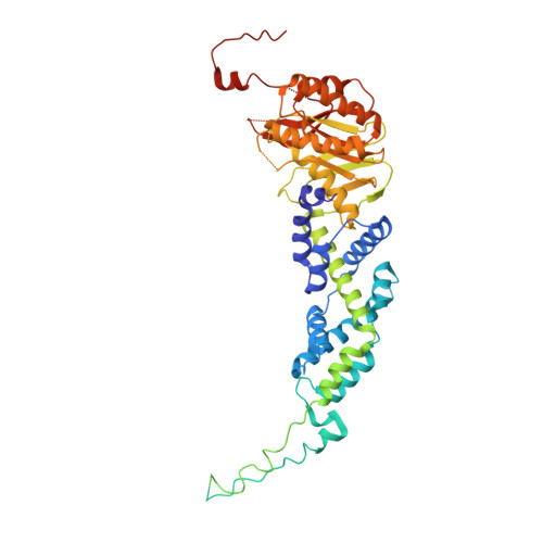
Crystal Structure of the core region of Thermus thermophilus MutS2 complexed with ADP.


Experimental Data Snapshot

  • Method: X-RAY DIFFRACTION
  • Resolution: 3.38 Å
  • R-Value Free: 0.272 
  • R-Value Work: 0.267 
  • R-Value Observed: 0.267 

wwPDB Validation   3D Report Full Report


Ligand Structure Quality Assessment 


This is version 1.2 of the entry. See complete history


Literature

Structural and functional insights into the mechanism by which MutS2 recognizes a DNA junction.

Fukui, K.Inoue, M.Murakawa, T.Baba, S.Kumasaka, T.Yano, T.

(2022) Structure 30: 973-982.e4

  • DOI: https://doi.org/10.1016/j.str.2022.03.014
  • Primary Citation of Related Structures:  
    7VUF, 7VUK

  • PubMed Abstract: 

    MutS family proteins are classified into MutS-I and -II lineages: MutS-I recognizes mismatched DNA and initiates mismatch repair, whereas MutS-II recognizes DNA junctions to modulate recombination. MutS-I forms dimeric clamp-like structures enclosing the mismatched DNA, and its composite ATPase sites regulate DNA-binding modes. Meanwhile, the structures of MutS-II have not been determined; accordingly, it remains unknown how MutS-II recognizes DNA junctions and how nucleotides control DNA binding. Here, we solved the ligand-free and ADP-bound crystal structures of bacterial MutS2 belonging to MutS-II. MutS2 also formed a dimeric clamp-like structure with composite ATPase sites. The ADP-bound MutS2 was more flexible compared to the ligand-free form and could be more suitable for DNA entry. The inner hole of the MutS2 clamp was two times larger than that of MutS-I, and site-directed mutagenesis analyses revealed DNA-binding sites at the inner hole. Based on these, a model is proposed that describes how MutS2 recognizes DNA junctions.


  • Organizational Affiliation

    Department of Biochemistry, Faculty of Medicine, Osaka Medical and Pharmaceutical University, Takatsuki, Osaka 569-8686, Japan. Electronic address: kenji.fukui@ompu.ac.jp.


Macromolecules
Find similar proteins by:  (by identity cutoff)  |  3D Structure
Entity ID: 1
MoleculeChains Sequence LengthOrganismDetailsImage
Endonuclease MutS2
A, B
488Thermus thermophilus HB8Mutation(s): 0 
Gene Names: mutS2TTHA1645
EC: 3.1
UniProt
Find proteins for Q5SHT5 (Thermus thermophilus (strain ATCC 27634 / DSM 579 / HB8))
Explore Q5SHT5 
Go to UniProtKB:  Q5SHT5
Entity Groups  
Sequence Clusters30% Identity50% Identity70% Identity90% Identity95% Identity100% Identity
UniProt GroupQ5SHT5
Sequence Annotations
Expand
  • Reference Sequence
Small Molecules
Ligands 5 Unique
IDChains Name / Formula / InChI Key2D Diagram3D Interactions
ADP (Subject of Investigation/LOI)
Query on ADP

Download Ideal Coordinates CCD File 
DA [auth B],
S [auth A]
ADENOSINE-5'-DIPHOSPHATE
C10 H15 N5 O10 P2
XTWYTFMLZFPYCI-KQYNXXCUSA-N
PEG
Query on PEG

Download Ideal Coordinates CCD File 
AA [auth B]
BA [auth B]
CA [auth B]
M [auth A]
N [auth A]
AA [auth B],
BA [auth B],
CA [auth B],
M [auth A],
N [auth A],
O [auth A],
P [auth A],
Q [auth A],
R [auth A],
Z [auth B]
DI(HYDROXYETHYL)ETHER
C4 H10 O3
MTHSVFCYNBDYFN-UHFFFAOYSA-N
CL
Query on CL

Download Ideal Coordinates CCD File 
K [auth A],
L [auth A],
Y [auth B]
CHLORIDE ION
Cl
VEXZGXHMUGYJMC-UHFFFAOYSA-M
MG
Query on MG

Download Ideal Coordinates CCD File 
C [auth A],
D [auth A],
E [auth A],
T [auth B],
U [auth B]
MAGNESIUM ION
Mg
JLVVSXFLKOJNIY-UHFFFAOYSA-N
NA
Query on NA

Download Ideal Coordinates CCD File 
F [auth A]
G [auth A]
H [auth A]
I [auth A]
J [auth A]
F [auth A],
G [auth A],
H [auth A],
I [auth A],
J [auth A],
V [auth B],
W [auth B],
X [auth B]
SODIUM ION
Na
FKNQFGJONOIPTF-UHFFFAOYSA-N
Experimental Data & Validation

Experimental Data

  • Method: X-RAY DIFFRACTION
  • Resolution: 3.38 Å
  • R-Value Free: 0.272 
  • R-Value Work: 0.267 
  • R-Value Observed: 0.267 
  • Space Group: P 41 21 2
Unit Cell:
Length ( Å )Angle ( ˚ )
a = 133.654α = 90
b = 133.654β = 90
c = 197.461γ = 90
Software Package:
Software NamePurpose
PHENIXrefinement
HKL-2000data reduction
HKL-2000data scaling
PDB_EXTRACTdata extraction
PHENIXphasing
PHENIXmodel building
Cootmodel building

Structure Validation

View Full Validation Report


Ligand Structure Quality Assessment 


Entry History & Funding Information

Deposition Data


Funding OrganizationLocationGrant Number
Not funded--

Revision History  (Full details and data files)

  • Version 1.0: 2022-04-06
    Type: Initial release
  • Version 1.1: 2022-10-19
    Changes: Database references
  • Version 1.2: 2023-11-29
    Changes: Data collection, Refinement description