7VUF

Crystal Structure of the core region of Thermus thermophilus MutS2.


Experimental Data Snapshot

  • Method: X-RAY DIFFRACTION
  • Resolution: 3.11 Å
  • R-Value Free: 0.279 
  • R-Value Work: 0.225 
  • R-Value Observed: 0.227 

wwPDB Validation   3D Report Full Report


This is version 1.1 of the entry. See complete history


Literature

Structural and functional insights into the mechanism by which MutS2 recognizes a DNA junction.

Fukui, K.Inoue, M.Murakawa, T.Baba, S.Kumasaka, T.Yano, T.

(2022) Structure 30: 973-982.e4

  • DOI: https://doi.org/10.1016/j.str.2022.03.014
  • Primary Citation of Related Structures:  
    7VUF, 7VUK

  • PubMed Abstract: 

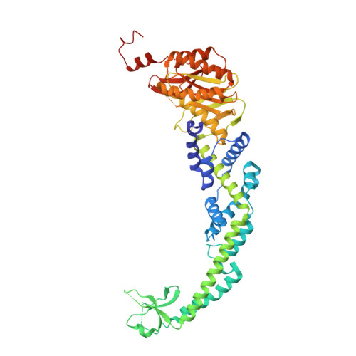
    MutS family proteins are classified into MutS-I and -II lineages: MutS-I recognizes mismatched DNA and initiates mismatch repair, whereas MutS-II recognizes DNA junctions to modulate recombination. MutS-I forms dimeric clamp-like structures enclosing the mismatched DNA, and its composite ATPase sites regulate DNA-binding modes. Meanwhile, the structures of MutS-II have not been determined; accordingly, it remains unknown how MutS-II recognizes DNA junctions and how nucleotides control DNA binding. Here, we solved the ligand-free and ADP-bound crystal structures of bacterial MutS2 belonging to MutS-II. MutS2 also formed a dimeric clamp-like structure with composite ATPase sites. The ADP-bound MutS2 was more flexible compared to the ligand-free form and could be more suitable for DNA entry. The inner hole of the MutS2 clamp was two times larger than that of MutS-I, and site-directed mutagenesis analyses revealed DNA-binding sites at the inner hole. Based on these, a model is proposed that describes how MutS2 recognizes DNA junctions.


  • Organizational Affiliation

    Department of Biochemistry, Faculty of Medicine, Osaka Medical and Pharmaceutical University, Takatsuki, Osaka 569-8686, Japan. Electronic address: kenji.fukui@ompu.ac.jp.


Macromolecules
Find similar proteins by:  (by identity cutoff)  |  3D Structure
Entity ID: 1
MoleculeChains Sequence LengthOrganismDetailsImage
Endonuclease MutS2A [auth B],
B [auth A],
C,
D
488Thermus thermophilus HB8Mutation(s): 0 
Gene Names: mutS2TTHA1645
EC: 3.1
UniProt
Find proteins for Q5SHT5 (Thermus thermophilus (strain ATCC 27634 / DSM 579 / HB8))
Explore Q5SHT5 
Go to UniProtKB:  Q5SHT5
Entity Groups  
Sequence Clusters30% Identity50% Identity70% Identity90% Identity95% Identity100% Identity
UniProt GroupQ5SHT5
Sequence Annotations
Expand
  • Reference Sequence
Experimental Data & Validation

Experimental Data

  • Method: X-RAY DIFFRACTION
  • Resolution: 3.11 Å
  • R-Value Free: 0.279 
  • R-Value Work: 0.225 
  • R-Value Observed: 0.227 
  • Space Group: P 21 21 21
Unit Cell:
Length ( Å )Angle ( ˚ )
a = 93.485α = 90
b = 114.207β = 90
c = 292.879γ = 90
Software Package:
Software NamePurpose
PHENIXrefinement
HKL-2000data reduction
HKL-2000data scaling
PDB_EXTRACTdata extraction
PHASERphasing
PHENIXmodel building
Cootmodel building

Structure Validation

View Full Validation Report


Entry History & Funding Information

Deposition Data


Funding OrganizationLocationGrant Number
Not funded--

Revision History  (Full details and data files)

  • Version 1.0: 2022-04-06
    Type: Initial release
  • Version 1.1: 2022-10-19
    Changes: Database references