7U6H | pdb_00007u6h

HalD with ornithine and alpha-ketoglutarate


Experimental Data Snapshot

  • Method: X-RAY DIFFRACTION
  • Resolution: 2.00 Å
  • R-Value Free: 
    0.213 (Depositor), 0.215 (DCC) 
  • R-Value Work: 
    0.177 (Depositor), 0.180 (DCC) 
  • R-Value Observed: 
    0.179 (Depositor) 

Starting Model: experimental
View more details

wwPDB Validation 3D Report Full Report

Validation slider image for 7U6H

Ligand Structure Quality Assessment 


This is version 1.2 of the entry. See complete history

Literature

Biocatalytic control of site-selectivity and chain length-selectivity in radical amino acid halogenases.

Kissman, E.N.Neugebauer, M.E.Sumida, K.H.Swenson, C.V.Sambold, N.A.Marchand, J.A.Millar, D.C.Chang, M.C.Y.

(2023) Proc Natl Acad Sci U S A 120: e2214512120-e2214512120

  • DOI: https://doi.org/10.1073/pnas.2214512120
  • Primary Citation Related Structures: 
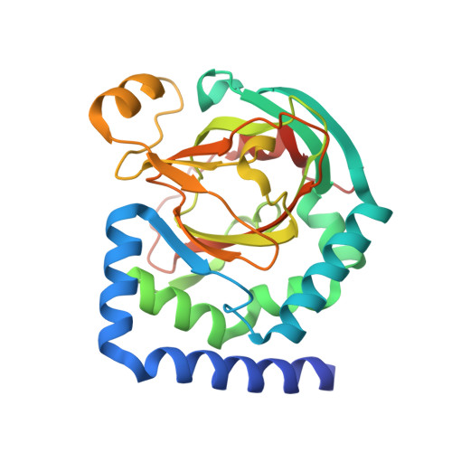
    7U6H, 7U6I, 7U6J

  • PubMed Abstract: 

    Biocatalytic C-H activation has the potential to merge enzymatic and synthetic strategies for bond formation. Fe II /αKG-dependent halogenases are particularly distinguished for their ability both to control selective C-H activation as well as to direct group transfer of a bound anion along a reaction axis separate from oxygen rebound, enabling the development of new transformations. In this context, we elucidate the basis for the selectivity of enzymes that perform selective halogenation to yield 4-Cl-lysine (BesD), 5-Cl-lysine (HalB), and 4-Cl-ornithine (HalD), allowing us to probe how site-selectivity and chain length selectivity are achieved. We now report the crystal structure of the HalB and HalD, revealing the key role of the substrate-binding lid in positioning the substrate for C 4 vs C 5 chlorination and recognition of lysine vs ornithine. Targeted engineering of the substrate-binding lid further demonstrates that these selectivities can be altered or switched, showcasing the potential to develop halogenases for biocatalytic applications.


  • Organizational Affiliation
    • Department of Chemistry, University of California, Berkeley, CA 94720.

Macromolecule Content 

  • Total Structure Weight: 126.28 kDa 
  • Atom Count: 9,369 
  • Modeled Residue Count: 1,012 
  • Deposited Residue Count: 1,056 
  • Unique protein chains: 1

Macromolecules

Find similar proteins by:|  3D Structure
Entity ID: 1
MoleculeChains  Sequence LengthOrganismDetailsImage
Halogenase D
A, B, C, D
264Pseudomonas kilonensisMutation(s): 0 
Gene Names: VP02_10785
UniProt
Find proteins for A0A0F4XRB2 (Pseudomonas kilonensis)
Explore A0A0F4XRB2 
Go to UniProtKB:  A0A0F4XRB2
Entity Groups
Sequence Clusters30% Identity50% Identity70% Identity90% Identity95% Identity100% Identity
UniProt GroupA0A0F4XRB2
Sequence Annotations
Expand
Reference Sequence

Small Molecules

Ligands 7 Unique
IDChains Name / Formula / InChI Key2D Diagram3D Interactions
PG4

Query on PG4


Download:Ideal Coordinates CCD File
O [auth B]TETRAETHYLENE GLYCOL
C8 H18 O5
UWHCKJMYHZGTIT-UHFFFAOYSA-N
PGE

Query on PGE


Download:Ideal Coordinates CCD File
DA [auth D]
EA [auth D]
H [auth A]
I [auth A]
J [auth A]
DA [auth D],
EA [auth D],
H [auth A],
I [auth A],
J [auth A],
R [auth B],
Y [auth C]
TRIETHYLENE GLYCOL
C6 H14 O4
ZIBGPFATKBEMQZ-UHFFFAOYSA-N
AKG
(Subject of Investigation/LOI)

Query on AKG


Download:Ideal Coordinates CCD File
FA [auth D],
K [auth A],
S [auth B],
Z [auth C]
2-OXOGLUTARIC ACID
C5 H6 O5
KPGXRSRHYNQIFN-UHFFFAOYSA-N
ORN
(Subject of Investigation/LOI)

Query on ORN


Download:Ideal Coordinates CCD File
CA [auth D],
G [auth A],
N [auth B],
V [auth C]
L-ornithine
C5 H12 N2 O2
AHLPHDHHMVZTML-BYPYZUCNSA-N
PEG

Query on PEG


Download:Ideal Coordinates CCD File
P [auth B],
Q [auth B],
W [auth C],
X [auth C]
DI(HYDROXYETHYL)ETHER
C4 H10 O3
MTHSVFCYNBDYFN-UHFFFAOYSA-N
NI

Query on NI


Download:Ideal Coordinates CCD File
AA [auth D],
E [auth A],
L [auth B],
T [auth C]
NICKEL (II) ION
Ni
VEQPNABPJHWNSG-UHFFFAOYSA-N
CL

Query on CL


Download:Ideal Coordinates CCD File
BA [auth D],
F [auth A],
M [auth B],
U [auth C]
CHLORIDE ION
Cl
VEXZGXHMUGYJMC-UHFFFAOYSA-M

Experimental Data & Validation

Experimental Data

  • Method: X-RAY DIFFRACTION
  • Resolution: 2.00 Å
  • R-Value Free:  0.213 (Depositor), 0.215 (DCC) 
  • R-Value Work:  0.177 (Depositor), 0.180 (DCC) 
  • R-Value Observed: 0.179 (Depositor) 
Space Group: P 1
Unit Cell:
Length ( Å )Angle ( ˚ )
a = 72.05α = 66.24
b = 73.226β = 75.8
c = 73.176γ = 85.28
Software Package:
Software NamePurpose
PHENIXrefinement
XDSdata reduction
Aimlessdata scaling
PHASERphasing
PDB_EXTRACTdata extraction

Structure Validation

View Full Validation Report


Ligand Structure Quality Assessment 


Entry History 

& Funding Information

Deposition Data


Funding OrganizationLocationGrant Number
National Institutes of Health/National Institute of General Medical Sciences (NIH/NIGMS)United States--

Revision History  (Full details and data files)

  • Version 1.0: 2023-03-15
    Type: Initial release
  • Version 1.1: 2023-03-22
    Changes: Database references
  • Version 1.2: 2023-10-25
    Changes: Data collection, Refinement description