7T1U

Crystal structure of a superbinder Src SH2 domain (sSrcF) in complex with a high affinity phosphopeptide


Experimental Data Snapshot

  • Method: X-RAY DIFFRACTION
  • Resolution: 2.65 Å
  • R-Value Free: 0.270 
  • R-Value Work: 0.218 
  • R-Value Observed: 0.224 

wwPDB Validation   3D Report Full Report


This is version 1.2 of the entry. See complete history


Literature

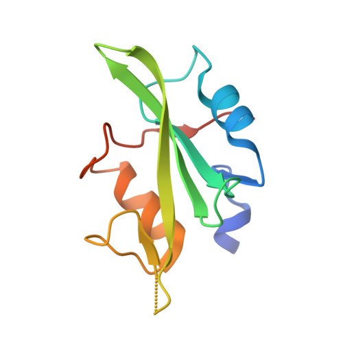
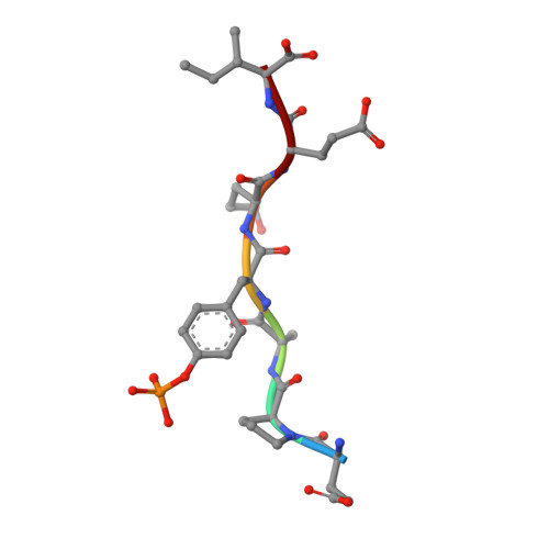
Engineered SH2 Domains for Targeted Phosphoproteomics.

Martyn, G.D.Veggiani, G.Kusebauch, U.Morrone, S.R.Yates, B.P.Singer, A.U.Tong, J.Manczyk, N.Gish, G.Sun, Z.Kurinov, I.Sicheri, F.Moran, M.F.Moritz, R.L.Sidhu, S.S.

(2022) ACS Chem Biol 17: 1472-1484

  • DOI: https://doi.org/10.1021/acschembio.2c00051
  • Primary Citation of Related Structures:  
    7T1K, 7T1L, 7T1U

  • PubMed Abstract: 

    A comprehensive analysis of the phosphoproteome is essential for understanding molecular mechanisms of human diseases. However, current tools used to enrich phosphotyrosine (pTyr) are limited in their applicability and scope. Here, we engineered new superbinder Src-Homology 2 (SH2) domains that enrich diverse sets of pTyr-peptides. We used phage display to select a Fes-SH2 domain variant (superFes; sFes 1 ) with high affinity for pTyr and solved its structure bound to a pTyr-peptide. We performed systematic structure-function analyses of the superbinding mechanisms of sFes 1 and superSrc-SH2 (sSrc 1 ), another SH2 superbinder. We grafted the superbinder motifs from sFes 1 and sSrc 1 into 17 additional SH2 domains and confirmed increased binding affinity for specific pTyr-peptides. Using mass spectrometry (MS), we demonstrated that SH2 superbinders have distinct specificity profiles and superior capabilities to enrich pTyr-peptides. Finally, using combinations of SH2 superbinders as affinity purification (AP) tools we showed that unique subsets of pTyr-peptides can be enriched with unparalleled depth and coverage.


  • Organizational Affiliation

    Donnelly Centre for Cellular and Biomolecular Research, University of Toronto, 160 College Street, Toronto, Ontario M5S3E1, Canada.


Macromolecules
Find similar proteins by:  (by identity cutoff)  |  3D Structure
Entity ID: 1
MoleculeChains Sequence LengthOrganismDetailsImage
Proto-oncogene tyrosine-protein kinase Src
A, B
111Homo sapiensMutation(s): 8 
Gene Names: SRCSRC1
EC: 2.7.10.2
UniProt & NIH Common Fund Data Resources
Find proteins for P12931 (Homo sapiens)
Explore P12931 
Go to UniProtKB:  P12931
PHAROS:  P12931
GTEx:  ENSG00000197122 
Entity Groups  
Sequence Clusters30% Identity50% Identity70% Identity90% Identity95% Identity100% Identity
UniProt GroupP12931
Sequence Annotations
Expand
  • Reference Sequence

Find similar proteins by:  Sequence   |   3D Structure  

Entity ID: 2
MoleculeChains Sequence LengthOrganismDetailsImage
Synthetic phosphopeptideC [auth D],
D [auth E]
7synthetic constructMutation(s): 0 
Sequence Annotations
Expand
  • Reference Sequence
Experimental Data & Validation

Experimental Data

  • Method: X-RAY DIFFRACTION
  • Resolution: 2.65 Å
  • R-Value Free: 0.270 
  • R-Value Work: 0.218 
  • R-Value Observed: 0.224 
  • Space Group: P 1
Unit Cell:
Length ( Å )Angle ( ˚ )
a = 34.87α = 75.85
b = 35.89β = 74.77
c = 53.98γ = 61.42
Software Package:
Software NamePurpose
PHENIXrefinement
MOSFLMdata reduction
Aimlessdata scaling
PHASERphasing

Structure Validation

View Full Validation Report


Entry History & Funding Information

Deposition Data


Funding OrganizationLocationGrant Number
Canadian Institutes of Health Research (CIHR)United StatesMOP-93684
Canadian Institutes of Health Research (CIHR)United StatesFDN-143277
National Institutes of Health/National Institute of General Medical Sciences (NIH/NIGMS)United StatesP30 GM124165

Revision History  (Full details and data files)

  • Version 1.0: 2022-08-24
    Type: Initial release
  • Version 1.1: 2023-10-18
    Changes: Data collection, Refinement description
  • Version 1.2: 2023-11-15
    Changes: Data collection