7SJ2

N-acetylglucosamine-1-phosphotransferase (GNPT) alpha and beta subunits (GNPTAB) catalytic domain, from zebrafish, in complex with uridine diphosphate N-acetylglucosamine (UDP-GlcNAc) and magnesium


Experimental Data Snapshot

  • Method: X-RAY DIFFRACTION
  • Resolution: 2.30 Å
  • R-Value Free: 0.202 
  • R-Value Work: 0.160 
  • R-Value Observed: 0.162 

wwPDB Validation   3D Report Full Report


Ligand Structure Quality Assessment 


This is version 1.2 of the entry. See complete history


Literature

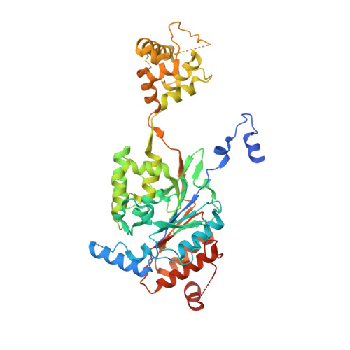
Structures of the mannose-6-phosphate pathway enzyme, GlcNAc-1-phosphotransferase.

Gorelik, A.Illes, K.Bui, K.H.Nagar, B.

(2022) Proc Natl Acad Sci U S A 119: e2203518119-e2203518119

  • DOI: https://doi.org/10.1073/pnas.2203518119
  • Primary Citation of Related Structures:  
    7S69, 7S6N, 7SJ2

  • PubMed Abstract: 

    The mannose-6-phosphate (M6P) pathway is responsible for the transport of hydrolytic enzymes to lysosomes. N-acetylglucosamine-1-phosphotransferase (GNPT) catalyzes the first step of tagging these hydrolases with M6P, which when recognized by receptors in the Golgi diverts them to lysosomes. Genetic defects in the GNPT subunits, GNPTAB and GNPTG, cause the lysosomal storage diseases mucolipidosis types II and III. To better understand its function, we determined partial three-dimensional structures of the GNPT complex. The catalytic domain contains a deep cavity for binding of uridine diphosphate- N -acetylglucosamine, and the surrounding residues point to a one-step transfer mechanism. An isolated structure of the gamma subunit of GNPT reveals that it can bind to mannose-containing glycans in different configurations, suggesting that it may play a role in directing glycans into the active site. These findings may facilitate the development of therapies for lysosomal storage diseases.


  • Organizational Affiliation

    Department of Biochemistry, McGill University, Montreal, QC H3G 0B1, Canada.


Macromolecules
Find similar proteins by:  (by identity cutoff)  |  3D Structure
Entity ID: 1
MoleculeChains Sequence LengthOrganismDetailsImage
N-acetylglucosamine-1-phosphotransferase subunit alpha,N-acetylglucosamine-1-phosphotransferase (GNPT) alpha (GNPTAB) catalytic domain,N-acetylglucosamine-1-phosphotransferase subunit beta
A, B
495Danio rerioDictyostelium discoideumMutation(s): 0 
Gene Names: gnptabgnptasi:ch211-234f20.3zgc:122985DDB0186697
UniProt
Find proteins for Q54MP1 (Dictyostelium discoideum)
Explore Q54MP1 
Go to UniProtKB:  Q54MP1
Find proteins for Q5RGJ8 (Danio rerio)
Explore Q5RGJ8 
Go to UniProtKB:  Q5RGJ8
Entity Groups  
Sequence Clusters30% Identity50% Identity70% Identity90% Identity95% Identity100% Identity
UniProt GroupsQ54MP1Q5RGJ8
Sequence Annotations
Expand
  • Reference Sequence
Oligosaccharides

Help

Entity ID: 2
MoleculeChains Length2D Diagram Glycosylation3D Interactions
alpha-D-mannopyranose-(1-3)-[alpha-D-mannopyranose-(1-6)]beta-D-mannopyranose-(1-4)-2-acetamido-2-deoxy-beta-D-glucopyranose-(1-4)-2-acetamido-2-deoxy-beta-D-glucopyranose
C
5N-Glycosylation
Glycosylation Resources
GlyTouCan:  G22768VO
GlyCosmos:  G22768VO
GlyGen:  G22768VO
Entity ID: 3
MoleculeChains Length2D Diagram Glycosylation3D Interactions
2-acetamido-2-deoxy-beta-D-glucopyranose-(1-4)-[alpha-L-fucopyranose-(1-6)]2-acetamido-2-deoxy-beta-D-glucopyranose
D, G
3N-Glycosylation
Glycosylation Resources
GlyTouCan:  G21290RB
GlyCosmos:  G21290RB
GlyGen:  G21290RB
Entity ID: 4
MoleculeChains Length2D Diagram Glycosylation3D Interactions
alpha-L-fucopyranose-(1-6)-2-acetamido-2-deoxy-beta-D-glucopyranose
E
2N-Glycosylation
Glycosylation Resources
GlyTouCan:  G86851RC
GlyCosmos:  G86851RC
GlyGen:  G86851RC
Entity ID: 5
MoleculeChains Length2D Diagram Glycosylation3D Interactions
beta-D-mannopyranose-(1-4)-2-acetamido-2-deoxy-beta-D-glucopyranose-(1-4)-2-acetamido-2-deoxy-beta-D-glucopyranose
F
3N-Glycosylation
Glycosylation Resources
GlyTouCan:  G15407YE
GlyCosmos:  G15407YE
GlyGen:  G15407YE
Small Molecules
Ligands 4 Unique
IDChains Name / Formula / InChI Key2D Diagram3D Interactions
UD1 (Subject of Investigation/LOI)
Query on UD1

Download Ideal Coordinates CCD File 
M [auth A],
S [auth B]
URIDINE-DIPHOSPHATE-N-ACETYLGLUCOSAMINE
C17 H27 N3 O17 P2
LFTYTUAZOPRMMI-CFRASDGPSA-N
NAG
Query on NAG

Download Ideal Coordinates CCD File 
H [auth A],
I [auth A],
N [auth B],
O [auth B]
2-acetamido-2-deoxy-beta-D-glucopyranose
C8 H15 N O6
OVRNDRQMDRJTHS-FMDGEEDCSA-N
CA
Query on CA

Download Ideal Coordinates CCD File 
J [auth A],
P [auth B]
CALCIUM ION
Ca
BHPQYMZQTOCNFJ-UHFFFAOYSA-N
MG (Subject of Investigation/LOI)
Query on MG

Download Ideal Coordinates CCD File 
K [auth A],
L [auth A],
Q [auth B],
R [auth B]
MAGNESIUM ION
Mg
JLVVSXFLKOJNIY-UHFFFAOYSA-N
Experimental Data & Validation

Experimental Data

  • Method: X-RAY DIFFRACTION
  • Resolution: 2.30 Å
  • R-Value Free: 0.202 
  • R-Value Work: 0.160 
  • R-Value Observed: 0.162 
  • Space Group: P 1 21 1
Unit Cell:
Length ( Å )Angle ( ˚ )
a = 94.542α = 90
b = 87.142β = 111.858
c = 108.369γ = 90
Software Package:
Software NamePurpose
PHENIXrefinement
HKL-2000data reduction
HKL-2000data scaling
PHASERphasing

Structure Validation

View Full Validation Report


Ligand Structure Quality Assessment 


Entry History & Funding Information

Deposition Data


Funding OrganizationLocationGrant Number
Canadian Institutes of Health Research (CIHR)CanadaPJT-173264

Revision History  (Full details and data files)

  • Version 1.0: 2022-05-25
    Type: Initial release
  • Version 1.1: 2022-08-17
    Changes: Database references
  • Version 1.2: 2023-10-18
    Changes: Data collection, Refinement description