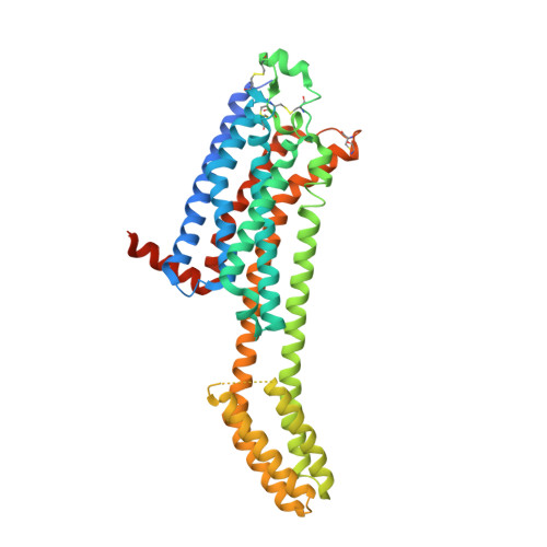
7RM5

MicroED structure of the human adenosine receptor at 2.8A


Experimental Data Snapshot

  • Method: ELECTRON CRYSTALLOGRAPHY
  • Resolution: 2.79 Å
  • R-Value Free: 0.288 
  • R-Value Work: 0.248 
  • R-Value Observed: 0.250 

wwPDB Validation   3D Report Full Report


This is version 1.3 of the entry. See complete history


Literature

MicroED structure of the human adenosine receptor determined from a single nanocrystal in LCP.

Martynowycz, M.W.Shiriaeva, A.Ge, X.Hattne, J.Nannenga, B.L.Cherezov, V.Gonen, T.

(2021) Proc Natl Acad Sci U S A 118

  • DOI: https://doi.org/10.1073/pnas.2106041118
  • Primary Citation of Related Structures:  
    7RM5

  • PubMed Abstract: 

    G protein-coupled receptors (GPCRs), or seven-transmembrane receptors, are a superfamily of membrane proteins that are critically important to physiological processes in the human body. Determining high-resolution structures of GPCRs without bound cognate signaling partners, such as a G protein, requires crystallization in lipidic cubic phase (LCP). GPCR crystals grown in LCP are often too small for traditional X-ray crystallography. These microcrystals are ideal for investigation by microcrystal electron diffraction (MicroED), but the gel-like nature of LCP makes traditional approaches to MicroED sample preparation insurmountable. Here, we show that the structure of a human A 2A adenosine receptor can be determined by MicroED after converting the LCP into the sponge phase followed by focused ion-beam milling. We determined the structure of the A 2A adenosine receptor to 2.8-Å resolution and resolved an antagonist in its orthosteric ligand-binding site, as well as four cholesterol molecules bound around the receptor. This study lays the groundwork for future structural studies of lipid-embedded membrane proteins by MicroED using single microcrystals that would be impossible with other crystallographic methods.


  • Organizational Affiliation

    HHMI, University of California, Los Angeles, CA 90095.


Macromolecules
Find similar proteins by:  (by identity cutoff)  |  3D Structure
Entity ID: 1
MoleculeChains Sequence LengthOrganismDetailsImage
Adenosine receptor A2a/Soluble cytochrome b562 chimera447Homo sapiensEscherichia coliMutation(s): 0 
Gene Names: ADORA2AADORA2cybC
Membrane Entity: Yes 
UniProt & NIH Common Fund Data Resources
Find proteins for P0ABE7 (Escherichia coli)
Explore P0ABE7 
Go to UniProtKB:  P0ABE7
Find proteins for P29274 (Homo sapiens)
Explore P29274 
Go to UniProtKB:  P29274
PHAROS:  P29274
GTEx:  ENSG00000128271 
Entity Groups  
Sequence Clusters30% Identity50% Identity70% Identity90% Identity95% Identity100% Identity
UniProt GroupsP0ABE7P29274
Sequence Annotations
Expand
  • Reference Sequence
Experimental Data & Validation

Experimental Data

  • Method: ELECTRON CRYSTALLOGRAPHY
  • Resolution: 2.79 Å
  • R-Value Free: 0.288 
  • R-Value Work: 0.248 
  • R-Value Observed: 0.250 
  • Space Group: C 2 2 21
Unit Cell:
Length ( Å )Angle ( ˚ )
a = 40.005α = 90
b = 180.54β = 90
c = 139.67γ = 90
Software Package:
Software NamePurpose
DIALSdata reduction
PHASERphasing
PHENIXrefinement
EM Software:
TaskSoftware PackageVersion
MODEL REFINEMENTPHENIX
RECONSTRUCTIONPHENIX

Structure Validation

View Full Validation Report


Entry History & Funding Information

Deposition Data


Funding OrganizationLocationGrant Number
Howard Hughes Medical Institute (HHMI)United States--
National Institutes of Health/National Institute of General Medical Sciences (NIH/NIGMS)United StatesP41GM136508
National Institutes of Health/National Institute of General Medical Sciences (NIH/NIGMS)United StatesR35 GM127086
National Institutes of Health/National Institute of General Medical Sciences (NIH/NIGMS)United StatesR01GM124152

Revision History  (Full details and data files)

  • Version 1.0: 2021-09-08
    Type: Initial release
  • Version 1.1: 2021-09-15
    Changes: Database references
  • Version 1.2: 2022-10-26
    Changes: Derived calculations
  • Version 1.3: 2023-10-18
    Changes: Data collection, Refinement description