7F1T

Crystal structure of the human chemokine receptor CCR5 in complex with MIP-1a


Experimental Data Snapshot

  • Method: X-RAY DIFFRACTION
  • Resolution: 2.60 Å
  • R-Value Free: 0.271 
  • R-Value Work: 0.228 

wwPDB Validation   3D Report Full Report


This is version 1.2 of the entry. See complete history


Literature

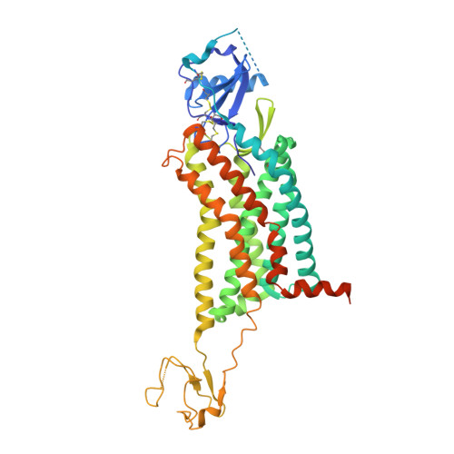
Structural basis for chemokine recognition and receptor activation of chemokine receptor CCR5.

Zhang, H.Chen, K.Tan, Q.Shao, Q.Han, S.Zhang, C.Yi, C.Chu, X.Zhu, Y.Xu, Y.Zhao, Q.Wu, B.

(2021) Nat Commun 12: 4151-4151

  • DOI: https://doi.org/10.1038/s41467-021-24438-5
  • Primary Citation of Related Structures:  
    7F1Q, 7F1R, 7F1S, 7F1T

  • PubMed Abstract: 

    The chemokine receptor CCR5 plays a vital role in immune surveillance and inflammation. However, molecular details that govern its endogenous chemokine recognition and receptor activation remain elusive. Here we report three cryo-electron microscopy structures of G i1 protein-coupled CCR5 in a ligand-free state and in complex with the chemokine MIP-1α or RANTES, as well as the crystal structure of MIP-1α-bound CCR5. These structures reveal distinct binding modes of the two chemokines and a specific accommodate pattern of the chemokine for the distal N terminus of CCR5. Together with functional data, the structures demonstrate that chemokine-induced rearrangement of toggle switch and plasticity of the receptor extracellular region are critical for receptor activation, while a conserved tryptophan residue in helix II acts as a trigger of receptor constitutive activation.


  • Organizational Affiliation

    CAS Key Laboratory of Receptor Research, Shanghai Institute of Materia Medica, Chinese Academy of Sciences, Shanghai, China.


Macromolecules
Find similar proteins by:  (by identity cutoff)  |  3D Structure
Entity ID: 1
MoleculeChains Sequence LengthOrganismDetailsImage
C-C motif chemokine 3,C-C chemokine receptor type 5,Rubredoxin,C-C chemokine receptor type 5491Homo sapiensClostridium pasteurianumMutation(s): 9 
Gene Names: CCL3G0S19-1MIP1ASCYA3CCR5CMKBR5
Membrane Entity: Yes 
UniProt & NIH Common Fund Data Resources
Find proteins for P51681 (Homo sapiens)
Explore P51681 
Go to UniProtKB:  P51681
PHAROS:  P51681
GTEx:  ENSG00000160791 
Find proteins for P10147 (Homo sapiens)
Explore P10147 
Go to UniProtKB:  P10147
PHAROS:  P10147
GTEx:  ENSG00000277632 
Find proteins for P00268 (Clostridium pasteurianum)
Explore P00268 
Go to UniProtKB:  P00268
Entity Groups  
Sequence Clusters30% Identity50% Identity70% Identity90% Identity95% Identity100% Identity
UniProt GroupsP00268P51681P10147
Sequence Annotations
Expand
  • Reference Sequence
Small Molecules
Ligands 1 Unique
IDChains Name / Formula / InChI Key2D Diagram3D Interactions
ZN
Query on ZN

Download Ideal Coordinates CCD File 
B [auth A]ZINC ION
Zn
PTFCDOFLOPIGGS-UHFFFAOYSA-N
Experimental Data & Validation

Experimental Data

  • Method: X-RAY DIFFRACTION
  • Resolution: 2.60 Å
  • R-Value Free: 0.271 
  • R-Value Work: 0.228 
  • Space Group: C 1 2 1
Unit Cell:
Length ( Å )Angle ( ˚ )
a = 49.133α = 90
b = 204.356β = 105.53
c = 69.012γ = 90
Software Package:
Software NamePurpose
BUSTERrefinement
HKL-2000data reduction
HKL-2000data scaling
PHASERphasing

Structure Validation

View Full Validation Report


Entry History & Funding Information

Deposition Data


Funding OrganizationLocationGrant Number
National Natural Science Foundation of China (NSFC)China31730027
National Natural Science Foundation of China (NSFC)China31825010

Revision History  (Full details and data files)

  • Version 1.0: 2021-07-14
    Type: Initial release
  • Version 1.1: 2021-07-28
    Changes: Database references
  • Version 1.2: 2023-11-29
    Changes: Data collection, Database references, Refinement description