7Z36

Crystal structure of the KAP1 tripartite motif in complex with the ZNF93 KRAB domain


Experimental Data Snapshot

  • Method: X-RAY DIFFRACTION
  • Resolution: 2.80 Å
  • R-Value Free: 0.274 
  • R-Value Work: 0.227 
  • R-Value Observed: 0.229 

wwPDB Validation   3D Report Full Report


This is version 1.2 of the entry. See complete history


Literature

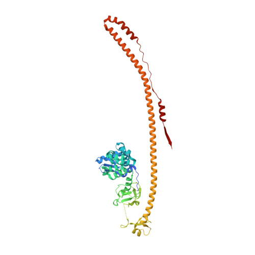
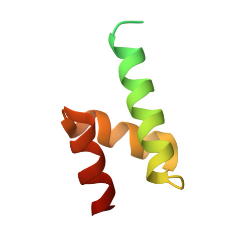
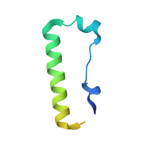
Structure and functional mapping of the KRAB-KAP1 repressor complex.

Stoll, G.A.Pandiloski, N.Douse, C.H.Modis, Y.

(2022) EMBO J 41: e111179-e111179

  • DOI: 10.15252/embj.2022111179
  • Primary Citation of Related Structures:  
    7Z36

  • PubMed Abstract: 
  • Transposable elements are a genetic reservoir from which new genes and regulatory elements can emerge. However, expression of transposable elements can be pathogenic and is therefore tightly controlled. KRAB domain-containing zinc finger proteins (KRAB-ZFPs) recruit the co-repressor KRAB-associated protein 1 (KAP1/TRIM28) to regulate many transposable elements, but how KRAB-ZFPs and KAP1 interact remains unclear ...

    Transposable elements are a genetic reservoir from which new genes and regulatory elements can emerge. However, expression of transposable elements can be pathogenic and is therefore tightly controlled. KRAB domain-containing zinc finger proteins (KRAB-ZFPs) recruit the co-repressor KRAB-associated protein 1 (KAP1/TRIM28) to regulate many transposable elements, but how KRAB-ZFPs and KAP1 interact remains unclear. Here, we report the crystal structure of the KAP1 tripartite motif (TRIM) in complex with the KRAB domain from a human KRAB-ZFP, ZNF93. Structure-guided mutations in the KAP1-KRAB binding interface abolished repressive activity in an epigenetic transcriptional silencing assay. Deposition of H3K9me3 over thousands of loci is lost genome-wide in cells expressing a KAP1 variant with mutations that abolish KRAB binding. Our work identifies and functionally validates the KRAB-KAP1 molecular interface, which is critical for a central transcriptional control axis in vertebrates. In addition, the structure-based prediction of KAP1 recruitment efficiency will enable optimization of KRABs used in CRISPRi.


    Organizational Affiliation

    Cambridge Institute of Therapeutic Immunology & Infectious Disease (CITIID), University of Cambridge School of Clinical Medicine, Cambridge, UK.


Macromolecules
Find similar proteins by:  (by identity cutoff)  |  3D Structure
Entity ID: 1
MoleculeChainsSequence LengthOrganismDetailsImage
Endolysin,Transcription intermediary factor 1-beta,Isoform 2 of Transcription intermediary factor 1-betaA, B482Homo sapiensMutation(s): 0 
Gene Names: TRIM28KAP1RNF96TIF1B
EC: 3.2.1.17 (PDB Primary Data), 2.3.2.27 (PDB Primary Data)
UniProt & NIH Common Fund Data Resources
Find proteins for Q13263-2 (Homo sapiens)
Explore Q13263-2 
Go to UniProtKB:  Q13263-2
Find proteins for P00720 (Enterobacteria phage T4)
Explore P00720 
Go to UniProtKB:  P00720
Find proteins for Q13263 (Homo sapiens)
Explore Q13263 
Go to UniProtKB:  Q13263
PHAROS:  Q13263
Entity Groups  
Sequence Clusters30% Identity50% Identity70% Identity90% Identity95% Identity100% Identity
UniProt GroupsQ13263Q13263-2P00720
Protein Feature View
Expand
  • Reference Sequence
Find similar proteins by:  (by identity cutoff)  |  3D Structure
Entity ID: 2
MoleculeChainsSequence LengthOrganismDetailsImage
SMARCAD1 CUE1 domainC,
E [auth S]
73Homo sapiensMutation(s): 0 
Entity Groups  
Sequence Clusters30% Identity50% Identity70% Identity90% Identity95% Identity100% Identity
Protein Feature View
Expand
  • Reference Sequence
Find similar proteins by:  (by identity cutoff)  |  3D Structure
Entity ID: 3
MoleculeChainsSequence LengthOrganismDetailsImage
Zinc finger protein 93D70Homo sapiensMutation(s): 0 
Gene Names: ZNF93ZNF505
UniProt & NIH Common Fund Data Resources
Find proteins for P35789 (Homo sapiens)
Explore P35789 
Go to UniProtKB:  P35789
PHAROS:  P35789
Entity Groups  
Sequence Clusters30% Identity50% Identity70% Identity90% Identity95% Identity100% Identity
UniProt GroupP35789
Protein Feature View
Expand
  • Reference Sequence
Experimental Data & Validation

Experimental Data

  • Method: X-RAY DIFFRACTION
  • Resolution: 2.80 Å
  • R-Value Free: 0.274 
  • R-Value Work: 0.227 
  • R-Value Observed: 0.229 
  • Space Group: C 1 2 1
  • Diffraction Data DOI: 10.15785/SBGRID/880
Unit Cell:
Length ( Å )Angle ( ˚ )
a = 190.283α = 90
b = 68.583β = 114.05
c = 149.111γ = 90
Software Package:
Software NamePurpose
xia2data reduction
PHENIXrefinement
DIALSdata scaling
Cootmodel building
PHASERphasing

Structure Validation

View Full Validation Report


Entry History & Funding Information

Deposition Data


Funding OrganizationLocationGrant Number
Medical Research Council (MRC, United Kingdom)United KingdomMR/S021604/1
Wellcome TrustUnited Kingdom205833/Z/16/Z

Revision History  (Full details and data files)

  • Version 1.0: 2022-11-02
    Type: Initial release
  • Version 1.1: 2022-11-23
    Changes: Database references
  • Version 1.2: 2022-12-28
    Changes: Database references