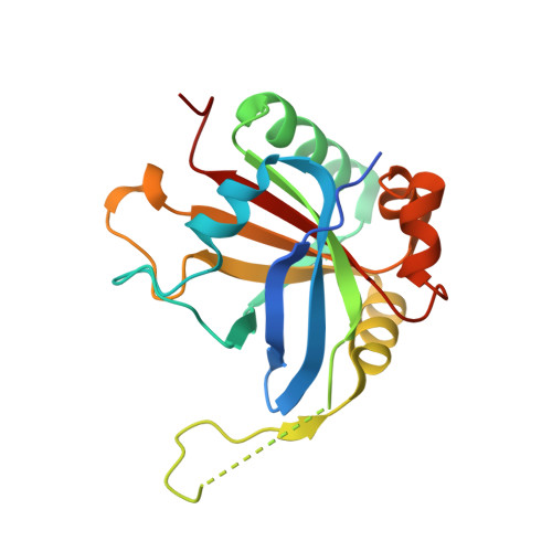
The SPOC domain is a phosphoserine binding module that bridges transcription machinery with co- and post-transcriptional regulators.
Appel, L.M., Franke, V., Benedum, J., Grishkovskaya, I., Strobl, X., Polyansky, A., Ammann, G., Platzer, S., Neudolt, A., Wunder, A., Walch, L., Kaiser, S., Zagrovic, B., Djinovic-Carugo, K., Akalin, A., Slade, D.(2023) Nat Commun 14: 166-166
- PubMed: 36631525
- DOI: https://doi.org/10.1038/s41467-023-35853-1
- Primary Citation of Related Structures:
7Z1K, 7Z27 - PubMed Abstract:
The heptad repeats of the C-terminal domain (CTD) of RNA polymerase II (Pol II) are extensively modified throughout the transcription cycle. The CTD coordinates RNA synthesis and processing by recruiting transcription regulators as well as RNA capping, splicing and 3'end processing factors. The SPOC domain of PHF3 was recently identified as a CTD reader domain specifically binding to phosphorylated serine-2 residues in adjacent CTD repeats. Here, we establish the SPOC domains of the human proteins DIDO, SHARP (also known as SPEN) and RBM15 as phosphoserine binding modules that can act as CTD readers but also recognize other phosphorylated binding partners. We report the crystal structure of SHARP SPOC in complex with CTD and identify the molecular determinants for its specific binding to phosphorylated serine-5. PHF3 and DIDO SPOC domains preferentially interact with the Pol II elongation complex, while RBM15 and SHARP SPOC domains engage with writers and readers of m 6 A, the most abundant RNA modification. RBM15 positively regulates m 6 A levels and mRNA stability in a SPOC-dependent manner, while SHARP SPOC is essential for its localization to inactive X-chromosomes. Our findings suggest that the SPOC domain is a major interface between the transcription machinery and regulators of transcription and co-transcriptional processes.
- Department of Radiation Oncology, Medical University of Vienna, Währinger Gürtel 18-20, 1090, Vienna, Austria.
Organizational Affiliation:


